 Explore
 Validate
 Learn
Learn Western blot
 ELISA
ELISAAntibody data
- Antibody Data
- Antigen structure
- References [0]
- Comments [0]
- Validations
- Western blot [2]
- Immunocytochemistry [1]
Submit
Validation data
Reference
Comment
Report error
- Product number
- MA1-21363 - Provider product page

- Provider
- Invitrogen Antibodies
- Product name
- FGF2 Monoclonal Antibody (F-343)
- Antibody type
- Monoclonal
- Antigen
- Purifed from natural sources
- Description
- MA1-21363 detects FGF Basic in Human samples.
- Reactivity
- Human
- Host
- Mouse
- Isotype
- IgG
- Antibody clone number
- F-343
- Vial size
- 250 µg
- Concentration
- 1 mg/mL
- Storage
- Store at 4°C short term. For long term storage, store at -20°C, avoiding freeze/thaw cycles.
No comments: Submit comment
Supportive validation
- Submitted by
- Invitrogen Antibodies (provider)
- Main image
- Experimental details
- Western blot analysis of recombinant human bFGF using anti-human bFGF monoclonal antibodies. From the left, third lane is FGF2 Monoclonal Antibody (F-343) (Product # MA1-21363).

- Submitted by
- Invitrogen Antibodies (provider)
- Main image

- Experimental details
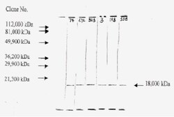
- Western blot was performed using Anti-FGF2 Monoclonal Antibody (F-343) (Product # MA1-21363) and 22kDa, 21kDa, 17kDa bands corresponding to FGF2 was observed in BJ, a fibroblast cell line, but not in T-47D and MCF7. Whole cell extracts (40 µg lysate) of BJ treated with PTI (1X for 4hrs) (Lane 1), BJ (Lane 2), T-47D treated with PTI (1X for 4hrs) (Lane 3), T-47D (Lane 4), MCF7 treated with PTI (1X for 4hrs) (Lane 5), MCF7 (Lane 6) were electrophoresed using NuPAGE™ 12% Bis-Tris Protein Gel (Product # NP0341BOX). Resolved proteins were then transferred onto a Nitrocellulose membrane (Product # IB23001) by iBlot® 2 Dry Blotting System (Product # IB21001). The blot was probed with the primary antibody (1:1250 dilution) and detected by chemiluminescence with Goat anti-Mouse IgG (H+L) Superclonal™ Recombinant Secondary Antibody, HRP (Product # A28177,1:8000 dilution) using the iBright FL 1000 (Product # A32752). Chemiluminescent detection was performed using Novex® ECL Chemiluminescent Substrate Reagent Kit (Product # WP20005). FGF2, a protein of the fibroblast growth factor family, is secreted in high abundance by fibroblast cell lines like BJ. Entrapment of FGF2 using PTI increases intracellular accumulation in BJ but not in T-47D and MCF7 [10.1158/1078-0432.CCR-10-2727].

Supportive validation
- Submitted by
- Invitrogen Antibodies (provider)
- Main image

- Experimental details
- Immunofluorescence analysis of FGF2 was performed using 80% confluent log phase BJ and T-47D control and PTI treated (1X for 4 hours) cells. The cells were fixed with 4% paraformaldehyde for 10 mins, permeabilized with 0.1% Triton™ X-100 for 10 mins, and blocked with 2% BSA for 45 mins at room temperature. The cells were labeled with FGF2 Monoclonal Antibody (F-343) (Product # MA1-21363) at 1:100 in 0.1% BSA, incubated at 4 degree celsius overnight and then labeled with Donkey anti-Mouse IgG (H+L) Highly Cross-Adsorbed Secondary Antibody, Alexa Fluor Plus 488 (Product # A32766), (1:2500 dilution), for 45 mins at room temperature (Panel a,e,i: Green). Nuclei (Panel b,f,j: Blue) were stained with ProLong™ Diamond Antifade Mountant with DAPI (Product # P36962). F-actin (Panel c,g,k: Red) was stained with Rhodamine Phalloidin (Product # R415, 1:300). Panel d represents the merged image of untreated BJ cells showing faint staining for FGF2 that is enhanced upon PTI treatment (Panel h). Panel i represents T-47D cells showing no staining for FGF2 upon PTI treatment. The images were captured at 60X magnification. FGF2 follows type I unconventional secretory pathway where it undergoes self-oligomerization and interacts with the inner and outer leaflets of the plasma membrane for its release. The punctate pattern of FGF2 seen in panel d and h is similar to that shown by Steringer et.al. of the intermediates and oligomers of FGF2 for membrane insertion.
