 Explore
 Validate
 Learn
Learn Western blot
 Immunohistochemistry
Antibody data
- Antibody Data
- Antigen structure
- References [2]
- Comments [0]
- Validations
- Immunohistochemistry [4]
Submit
Validation data
Reference
Comment
Report error
- Product number
- NB600-1536 - Provider product page

- Provider
- Novus Biologicals
- Proper citation
- Novus Cat#NB600-1536, RRID:AB_789874
- Product name
- Rabbit Polyclonal FGF basic/FGF2/bFGF Antibody
- Antibody type
- Polyclonal
- Description
- Sterile filtered. Does not cross react with bovine FGF Acidic or bovine serum albumin.
- Reactivity
- Human, Rat, Bovine
- Host
- Rabbit
- Isotype
- IgG
- Vial size
- 0.1 ml
- Concentration
- LYOPH
- Storage
- Store at 4C short term. Aliquot and store at -20C long term. Avoid freeze-thaw cycles.
Submitted references Renal heparan sulfate proteoglycans modulate fibroblast growth factor 2 signaling in experimental chronic transplant dysfunction.
Fibroblast growth factor-2 expression in the preimplantation equine conceptus and endometrium of pregnant and cyclic mares.
Katta K, Boersema M, Adepu S, Rienstra H, Celie JWAM, Mencke R, Molema G, van Goor H, Berden JHM, Navis G, Hillebrands JL, van den Born J
The American journal of pathology 2013 Nov;183(5):1571-1584
The American journal of pathology 2013 Nov;183(5):1571-1584
Fibroblast growth factor-2 expression in the preimplantation equine conceptus and endometrium of pregnant and cyclic mares.
de Ruijter-Villani M, van Boxtel PR, Stout TA
Theriogenology 2013 Dec;80(9):979-89
Theriogenology 2013 Dec;80(9):979-89
No comments: Submit comment
Supportive validation
- Submitted by
- Novus Biologicals (provider)
- Main image
- Experimental details
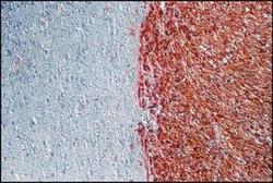
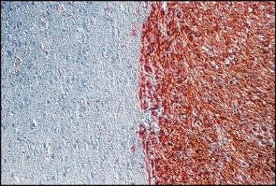
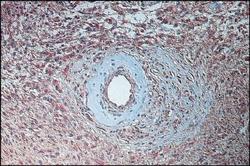
- Immunohistochemistry: FGF basic/FGF2 Antibody [NB600-1536] - Immunoperoxidase staining (PAP) of U87MG glioma cell suspension implanted into nude rat brain (Han rnu/rnu) using Rabbit Anti-Fibroblast Growth Factor-Basic (NB600-1536). High bFGF expression is exclusively shown within tumor endothelial cells. The tissue sample was paraffin-embedded and 4% Tris-buffered formalin-fixed without any enzymatic or antigen-demasking pre-processing. From Dr. A.-C. Stan, Institute of Neuropathology, Medical School Hannover, Hannover, Germany. Ref: Stan, A.C., et al., J. Neurosurg., 82, 1044 (1995).
- Submitted by
- Novus Biologicals (provider)
- Main image
- Experimental details
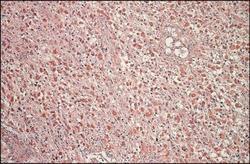
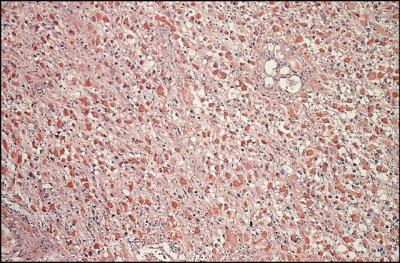
- Immunohistochemistry: FGF basic/FGF2 Antibody [NB600-1536] - Immunoperoxidase staining (PAP) of a gemistocytic differentiated area of an anaplastic astrocytoma (WHO grade III, SAMS grade 3) using Rabbit Anti-Fibroblast Growth Factor-Basic. High bFGF expression is shown within tumor cells as well as within the endothelial cells.
- Submitted by
- Novus Biologicals (provider)
- Main image

- Experimental details
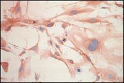
- Immunohistochemistry: FGF basic/FGF2 Antibody [NB600-1536] - Immunoperoxidase staining (PAP) of a freshly explanted gliobastoma multiforme (WHO grade IV, SAMS grade 4) in culture (passage # 3) using Rabbit Anti-Fibroblast Growth Factor-Basic (NB600-1356). Intracytoplasmatically, homogeneous, high bFGF expression is shown within highly polymorphic tumor cells. The tumor cells on glass coverslips were methanol/acetone (100%, ice-cold, 1:1) fixed without any enzymatic or antigen-demasking pre-processing. From Dr. A.-C. Stan, Institute of Neuropathology, Medical School Hannover, Hannover, Germany.
- Submitted by
- Novus Biologicals (provider)
- Main image
- Experimental details
- Immunohistochemistry: FGF basic/FGF2 Antibody [NB600-1536] - Immunoperoxidase staining (PAP) of a fibrillary differentiated astrocytoma (WHO grade I, SAMS grade 1) using Rabbit Anti-Fibroblast Growth Factor-Basic. High bFGF expression is shown within tumor cells as well as within endothelial cells .
