 Explore
 Validate
 Learn
Learn Western blot
 ELISA
ELISA Immunocytochemistry
Antibody data
- Antibody Data
- Antigen structure
- References [1]
- Comments [0]
- Validations
- Immunocytochemistry [2]
- Immunohistochemistry [1]
Submit
Validation data
Reference
Comment
Report error
- Product number
- MA5-15276 - Provider product page

- Provider
- Invitrogen Antibodies
- Product name
- FGF2 Monoclonal Antibody (2H5G2C11)
- Antibody type
- Monoclonal
- Antigen
- Purifed from natural sources
- Description
- MA5-15276 targets FGF2 in indirect ELISA, IHC and WB applications and shows reactivity with Human samples. The MA5-15276 immunogen is purified recombinant fragment of FGF2 expressed in E. Coli.
- Reactivity
- Human
- Host
- Mouse
- Isotype
- IgG
- Antibody clone number
- 2H5G2C11
- Vial size
- 100 μL
- Concentration
- Conc. Not Determined
- Storage
- Store at 4°C short term. For long term storage, store at -20°C, avoiding freeze/thaw cycles.
Submitted references c-Myc-mediated repression of miR-15-16 in hypoxia is induced by increased HIF-2α and promotes tumor angiogenesis and metastasis by upregulating FGF2.
Xue G, Yan HL, Zhang Y, Hao LQ, Zhu XT, Mei Q, Sun SH
Oncogene 2015 Mar 12;34(11):1393-406
Oncogene 2015 Mar 12;34(11):1393-406
No comments: Submit comment
Supportive validation
- Submitted by
- Invitrogen Antibodies (provider)
- Main image

- Experimental details
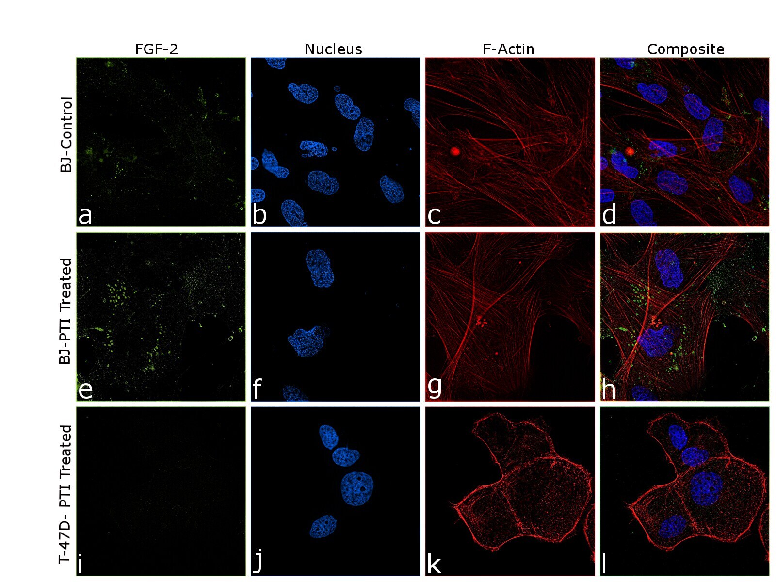
- Immunofluorescence analysis of FGF2 was performed using 80% confluent log phase BJ and T-47D, control and PTI treated, cells. The cells were fixed with 4% paraformaldehyde for 10 minutes, permeabilized with 0.1% Triton™ X-100 for 10 minutes, and blocked with 2% BSA for 45 minutes at room temperature. The cells were labeled with FGF2 Monoclonal Antibody (2H5G2C11) (Product # MA5-15276) at 1:100 in 0.1% BSA, incubated at 4 degree celsius overnight and then labeled with Donkey anti-Mouse IgG (H+L) Highly Cross-Adsorbed Secondary Antibody, Alexa Fluor Plus 488 (Product # A32766), (1:2500 dilution), for 45 minutes at room temperature (Panel a,e,i: Green). Nuclei (Panel b,f,j: Blue) were stained with ProLong™ Diamond Antifade Mountant with DAPI (Product # P36962). F-actin (Panel c,g,k: Red) was stained with Rhodamine Phalloidin (Product # R415, 1:300). Panel d represents the merged image of untreated BJ cells showing faint staining for FGF2 that is enhanced upon PTI treatment (Panel h). Panel l represents T-47D cells showing no staining for FGF2 upon PTI treatment. The images were captured at 60X magnification. FGF2 follows type I unconventional secretory pathway where it undergoes self-oligomerization and interacts with the inner and outer leaflets of the plasma membrane for its release. The punctate pattern of FGF2 seen in panel d and h is similar to that shown by Steringer et.al. of the intermediates and oligomers of FGF2 for membrane insertion.
- Submitted by
- Invitrogen Antibodies (provider)
- Main image

- Experimental details
- Immunofluorescence analysis of FGF2 was performed using 80% confluent log phase BJ and T-47D, control and PTI treated, cells. The cells were fixed with 4% paraformaldehyde for 10 minutes, permeabilized with 0.1% Triton™ X-100 for 10 minutes, and blocked with 2% BSA for 45 minutes at room temperature. The cells were labeled with FGF2 Monoclonal Antibody (2H5G2C11) (Product # MA5-15276) at 1:100 in 0.1% BSA, incubated at 4 degree celsius overnight and then labeled with Donkey anti-Mouse IgG (H+L) Highly Cross-Adsorbed Secondary Antibody, Alexa Fluor Plus 488 (Product # A32766), (1:2500 dilution), for 45 minutes at room temperature (Panel a,e,i: Green). Nuclei (Panel b,f,j: Blue) were stained with ProLong™ Diamond Antifade Mountant with DAPI (Product # P36962). F-actin (Panel c,g,k: Red) was stained with Rhodamine Phalloidin (Product # R415, 1:300). Panel d represents the merged image of untreated BJ cells showing faint staining for FGF2 that is enhanced upon PTI treatment (Panel h). Panel l represents T-47D cells showing no staining for FGF2 upon PTI treatment. The images were captured at 60X magnification. FGF2 follows type I unconventional secretory pathway where it undergoes self-oligomerization and interacts with the inner and outer leaflets of the plasma membrane for its release. The punctate pattern of FGF2 seen in panel d and h is similar to that shown by Steringer et.al. of the intermediates and oligomers of FGF2 for membrane insertion.

Supportive validation
- Submitted by
- Invitrogen Antibodies (provider)
- Main image
- Experimental details
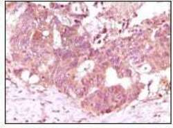
- Immunohistochemical analysis of paraffin-embedded human rectum adenocarcinoma tissue showing cytoplasmic localization using FGF2 monoclonal antibody (Product # MA5-15276) followed with DAB staining.
