 Explore
 Validate
 Learn
Learn Western blot
Antibody data
- Antibody Data
- Antigen structure
- References [0]
- Comments [0]
- Validations
- Western blot [5]
- Immunohistochemistry [2]
Submit
Validation data
Reference
Comment
Report error
- Product number
- LS-C100260 - Provider product page

- Provider
- LSBio
- Proper citation
- LifeSpan Cat#LS-C100260, RRID:AB_2277970
- Product name
- EPHA4 / EPH Receptor A4 Antibody (aa875-904) LS-C100260
- Antibody type
- Polyclonal
- Description
- Ammonium sulfate precipitation
- Reactivity
- Human
- Host
- Rabbit
- Storage
- Maintain refrigerated at 2°C to 8°C for up to 6 months. For long term storage store at -20°C.
No comments: Submit comment
Enhanced validation
- Submitted by
- LSBio (provider)
- Enhanced method
- Genetic validation
- Main image

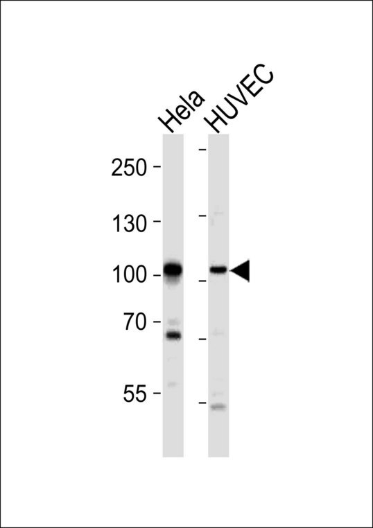
- Experimental details
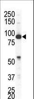
- Western blot of lysates from HeLa, HUVEC cell line (from left to right), using EPHA4 Antibody (R890). Antibody was diluted at 1:1000 at each lane. A goat anti-rabbit IgG H&L (HRP) at 1:10000 dilution was used as the secondary antibody. Lysates at 20ug per lane.
- Submitted by
- LSBio (provider)
- Enhanced method
- Genetic validation
- Main image
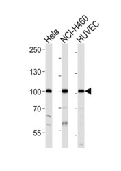
- Experimental details
- Western blot of lysates from HeLa, NCI-H460, HUVEC cell line (from left to right), using EPHA4 Antibody (R890). Antibody was diluted at 1:1000 at each lane. A goat anti-rabbit IgG H&L (HRP) at 1:10000 dilution was used as the secondary antibody. Lysates at 20ug per lane.

- Submitted by
- LSBio (provider)
- Enhanced method
- Genetic validation
- Main image
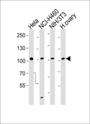
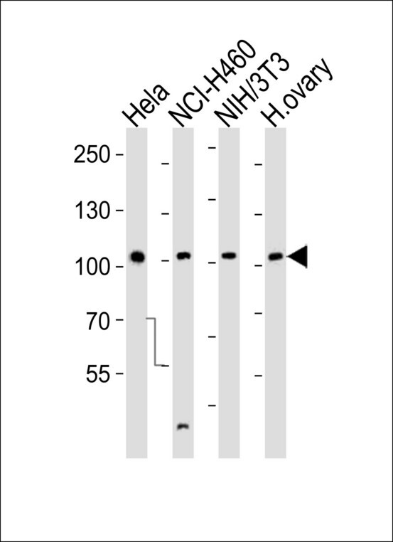
- Experimental details
- Western blot of lysates from HeLa, NCI-H460, mouse NIH/3T3 cell line and human ovary tissue lysate (from left to right), using EPHA4 Antibody (R890). Antibody was diluted at 1:1000 at each lane. A goat anti-rabbit IgG H&L (HRP) at 1:10000 dilution was used as the secondary antibody. Lysates at 35ug per lane.
- Submitted by
- LSBio (provider)
- Enhanced method
- Genetic validation
- Main image
- Experimental details
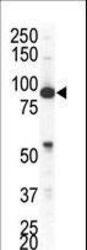
- Western blot of anti-EphA4 antibody in NCI-H460 cell lysate. EphA4 (arrow) was detected using purified antibody. Secondary HRP-anti-rabbit was used for signal visualization with chemiluminescence.
- Submitted by
- LSBio (provider)
- Enhanced method
- Genetic validation
- Main image
- Experimental details
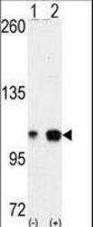
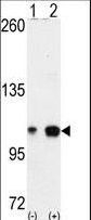
- Western blot of EPHA4 (arrow) using EphA4 Antibody. 293 cell lysates (2 ug/lane) either nontransfected (Lane 1) or transiently transfected with the EPHA4 gene (Lane 2) (Origene Technologies).
Enhanced validation
- Submitted by
- LSBio (provider)
- Enhanced method
- Genetic validation
- Main image

- Experimental details
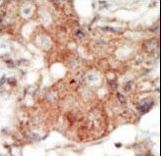
- Formalin-fixed and paraffin-embedded human cancer tissue reacted with the primary antibody, which was peroxidase-conjugated to the secondary antibody, followed by DAB staining. This data demonstrates the use of this antibody for immunohistochemistry; clinical relevance has not been evaluated. BC = breast carcinoma; HC = hepatocarcinoma.
- Submitted by
- LSBio (provider)
- Main image
- Experimental details
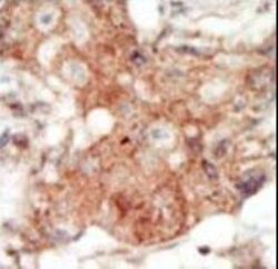
- Formalin-fixed and paraffin-embedded human cancer tissue reacted with the primary antibody, which was peroxidase-conjugated to the secondary antibody, followed by DAB staining. This data demonstrates the use of this antibody for immunohistochemistry; clinical relevance has not been evaluated. BC = breast carcinoma; HC = hepatocarcinoma.
