Explore
 Validate
 Learn
Learn Western blot
 Other assay
Antibody data
- Antibody Data
- Antigen structure
- References [22]
- Comments [0]
- Validations
- Other assay [14]
Submit
Validation data
Reference
Comment
Report error
- Product number
- 37-1600 - Provider product page

- Provider
- Invitrogen Antibodies
- Product name
- EphA4 Monoclonal Antibody (4C8H5)
- Antibody type
- Monoclonal
- Antigen
- Synthetic peptide
- Description
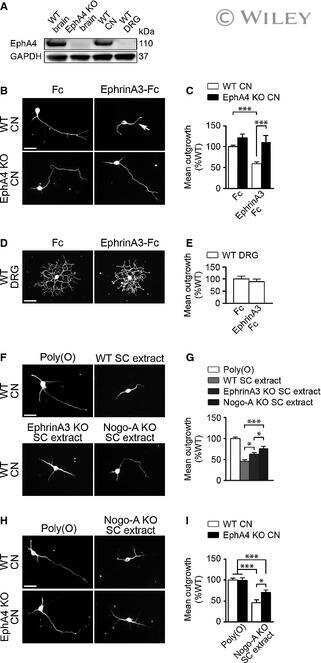
- This antibody is specific for the endogenous EphA4 receptor protein. No cross-reactivity with EphA5 or EphA7 has been detected. On western blots, this antibody identifies a band at ~110-120 kDa. Reactivity has been confirmed with EphA4-transfected 293T cells and mouse brain lysates by western blot and immunoprecipitation and on transfected 293 cells and mouse hippocampus by immunohistochemistry.
- Reactivity
- Human, Mouse, Chicken/Avian
- Host
- Mouse
- Isotype
- IgG
- Antibody clone number
- 4C8H5
- Vial size
- 100 μg
- Concentration
- 0.5 mg/mL
- Storage
- -20°C
Submitted references EphA4 targeting agents protect motor neurons from cell death induced by amyotrophic lateral sclerosis -astrocytes.
NMR-Guided Design of Potent and Selective EphA4 Agonistic Ligands.
Environmental enrichment during the chronic phase after experimental stroke promotes functional recovery without synergistic effects of EphA4 targeted therapy.
Lowering EphA4 Does Not Ameliorate Disease in a Mouse Model for Severe Spinal Muscular Atrophy.
Blood vessels guide Schwann cell migration in the adult demyelinated CNS through Eph/ephrin signaling.
Reducing EphA4 before disease onset does not affect survival in a mouse model of Amyotrophic Lateral Sclerosis.
Peripheral loss of EphA4 ameliorates TBI-induced neuroinflammation and tissue damage.
EphA4 loss improves social memory performance and alters dendritic spine morphology without changes in amyloid pathology in a mouse model of Alzheimer's disease.
Ephrin receptor A2 is a functional entry receptor for Epstein-Barr virus.
Potent and Selective EphA4 Agonists for the Treatment of ALS.
An Epha4/Sipa1l3/Wnt pathway regulates eye development and lens maturation.
Eph receptor interclass cooperation is required for the regulation of cell proliferation.
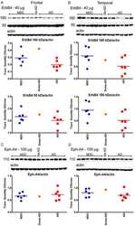
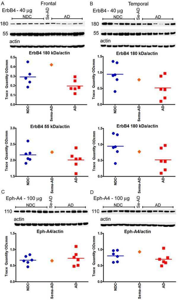
Neuropathological and biochemical assessments of an Alzheimer's disease patient treated with the γ-secretase inhibitor semagacestat.
The Ephrin receptor EphA4 restricts axonal sprouting and enhances branching in the injured mouse optic nerve.
Modifying expression of EphA4 and its downstream targets improves functional recovery after stroke.
Upregulation of axon guidance molecules in the adult central nervous system of Nogo-A knockout mice restricts neuronal growth and regeneration.
Activation of EphA receptors mediates the recruitment of the adaptor protein Slap, contributing to the downregulation of N-methyl-D-aspartate receptors.
EPHA4 is a disease modifier of amyotrophic lateral sclerosis in animal models and in humans.
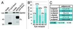
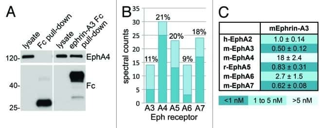
Profiling Eph receptor expression in cells and tissues: a targeted mass spectrometry approach.
Meltrin beta/ADAM19 interacting with EphA4 in developing neural cells participates in formation of the neuromuscular junction.
alpha2-Chimaerin is an essential EphA4 effector in the assembly of neuronal locomotor circuits.
The EphA4 receptor regulates neuronal morphology through SPAR-mediated inactivation of Rap GTPases.
Dennys C, Baggio C, Rodrigo R, Roussel F, Kulinich A, Heintzman S, Fox A, Kolb SJ, Shaw PJ, Ethell IM, Pellecchia M, Meyer KC
iScience 2022 Sep 16;25(9):104877
iScience 2022 Sep 16;25(9):104877
NMR-Guided Design of Potent and Selective EphA4 Agonistic Ligands.
Baggio C, Kulinich A, Dennys CN, Rodrigo R, Meyer K, Ethell I, Pellecchia M
Journal of medicinal chemistry 2021 Aug 12;64(15):11229-11246
Journal of medicinal chemistry 2021 Aug 12;64(15):11229-11246
Environmental enrichment during the chronic phase after experimental stroke promotes functional recovery without synergistic effects of EphA4 targeted therapy.
de Boer A, Storm A, Gomez-Soler M, Smolders S, Rué L, Poppe L, B Pasquale E, Robberecht W, Lemmens R
Human molecular genetics 2020 Mar 13;29(4):605-617
Human molecular genetics 2020 Mar 13;29(4):605-617
Lowering EphA4 Does Not Ameliorate Disease in a Mouse Model for Severe Spinal Muscular Atrophy.
Poppe L, Smolders S, Rué L, Timmers M, Lenaerts A, Storm A, Schoonaert L, de Boer A, Van Damme P, Van Den Bosch L, Robberecht W, Lemmens R
Frontiers in neuroscience 2019;13:1233
Frontiers in neuroscience 2019;13:1233
Blood vessels guide Schwann cell migration in the adult demyelinated CNS through Eph/ephrin signaling.
Garcia-Diaz B, Bachelin C, Coulpier F, Gerschenfeld G, Deboux C, Zujovic V, Charnay P, Topilko P, Baron-Van Evercooren A
Acta neuropathologica 2019 Sep;138(3):457-476
Acta neuropathologica 2019 Sep;138(3):457-476
Reducing EphA4 before disease onset does not affect survival in a mouse model of Amyotrophic Lateral Sclerosis.
Rué L, Timmers M, Lenaerts A, Smolders S, Poppe L, de Boer A, Van Den Bosch L, Van Damme P, Robberecht W, Lemmens R
Scientific reports 2019 Oct 1;9(1):14112
Scientific reports 2019 Oct 1;9(1):14112
Peripheral loss of EphA4 ameliorates TBI-induced neuroinflammation and tissue damage.
Kowalski EA, Chen J, Hazy A, Fritsch LE, Gudenschwager-Basso EK, Chen M, Wang X, Qian Y, Zhou M, Byerly M, Pickrell AM, Matson JB, Allen IC, Theus MH
Journal of neuroinflammation 2019 Nov 11;16(1):210
Journal of neuroinflammation 2019 Nov 11;16(1):210
EphA4 loss improves social memory performance and alters dendritic spine morphology without changes in amyloid pathology in a mouse model of Alzheimer's disease.
Poppe L, Rué L, Timmers M, Lenaerts A, Storm A, Callaerts-Vegh Z, Courtand G, de Boer A, Smolders S, Van Damme P, Van Den Bosch L, D'Hooge R, De Strooper B, Robberecht W, Lemmens R
Alzheimer's research & therapy 2019 Dec 12;11(1):102
Alzheimer's research & therapy 2019 Dec 12;11(1):102
Ephrin receptor A2 is a functional entry receptor for Epstein-Barr virus.
Chen J, Sathiyamoorthy K, Zhang X, Schaller S, Perez White BE, Jardetzky TS, Longnecker R
Nature microbiology 2018 Feb;3(2):172-180
Nature microbiology 2018 Feb;3(2):172-180
Potent and Selective EphA4 Agonists for the Treatment of ALS.
Wu B, De SK, Kulinich A, Salem AF, Koeppen J, Wang R, Barile E, Wang S, Zhang D, Ethell I, Pellecchia M
Cell chemical biology 2017 Mar 16;24(3):293-305
Cell chemical biology 2017 Mar 16;24(3):293-305
An Epha4/Sipa1l3/Wnt pathway regulates eye development and lens maturation.
Rothe M, Kanwal N, Dietmann P, Seigfried FA, Hempel A, Schütz D, Reim D, Engels R, Linnemann A, Schmeisser MJ, Bockmann J, Kühl M, Boeckers TM, Kühl SJ
Development (Cambridge, England) 2017 Jan 15;144(2):321-333
Development (Cambridge, England) 2017 Jan 15;144(2):321-333
Eph receptor interclass cooperation is required for the regulation of cell proliferation.
Jurek A, Genander M, Kundu P, Catchpole T, He X, Strååt K, Sabelström H, Xu NJ, Pettersson S, Henkemeyer M, Frisén J
Experimental cell research 2016 Oct 15;348(1):10-22
Experimental cell research 2016 Oct 15;348(1):10-22
Neuropathological and biochemical assessments of an Alzheimer's disease patient treated with the γ-secretase inhibitor semagacestat.
Roher AE, Maarouf CL, Kokjohn TA, Whiteside CM, Kalback WM, Serrano G, Belden C, Liebsack C, Jacobson SA, Sabbagh MN, Beach TG
American journal of neurodegenerative disease 2014;3(3):115-33
American journal of neurodegenerative disease 2014;3(3):115-33
The Ephrin receptor EphA4 restricts axonal sprouting and enhances branching in the injured mouse optic nerve.
Joly S, Jordi N, Schwab ME, Pernet V
The European journal of neuroscience 2014 Oct;40(7):3021-31
The European journal of neuroscience 2014 Oct;40(7):3021-31
Modifying expression of EphA4 and its downstream targets improves functional recovery after stroke.
Lemmens R, Jaspers T, Robberecht W, Thijs VN
Human molecular genetics 2013 Jun 1;22(11):2214-20
Human molecular genetics 2013 Jun 1;22(11):2214-20
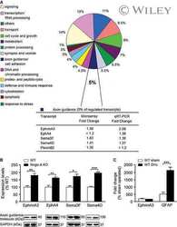
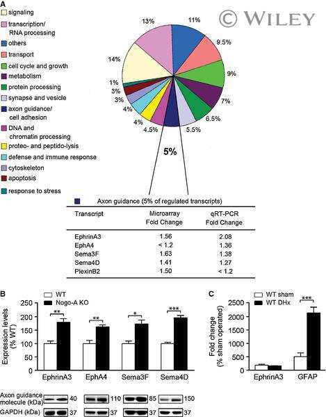
Upregulation of axon guidance molecules in the adult central nervous system of Nogo-A knockout mice restricts neuronal growth and regeneration.
Kempf A, Montani L, Petrinovic MM, Schroeter A, Weinmann O, Patrignani A, Schwab ME
The European journal of neuroscience 2013 Dec;38(11):3567-79
The European journal of neuroscience 2013 Dec;38(11):3567-79
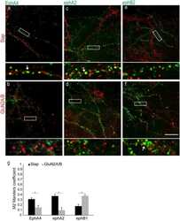
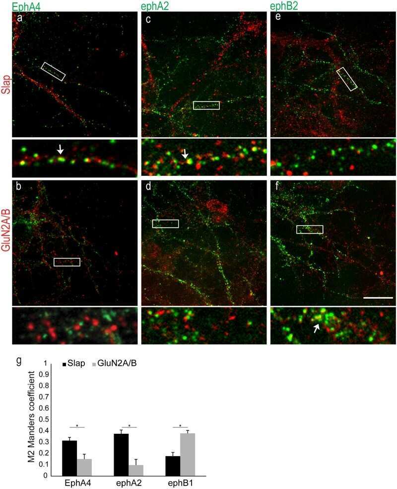
Activation of EphA receptors mediates the recruitment of the adaptor protein Slap, contributing to the downregulation of N-methyl-D-aspartate receptors.
Semerdjieva S, Abdul-Razak HH, Salim SS, Yáñez-Muñoz RJ, Chen PE, Tarabykin V, Alifragis P
Molecular and cellular biology 2013 Apr;33(7):1442-55
Molecular and cellular biology 2013 Apr;33(7):1442-55
EPHA4 is a disease modifier of amyotrophic lateral sclerosis in animal models and in humans.
Van Hoecke A, Schoonaert L, Lemmens R, Timmers M, Staats KA, Laird AS, Peeters E, Philips T, Goris A, Dubois B, Andersen PM, Al-Chalabi A, Thijs V, Turnley AM, van Vught PW, Veldink JH, Hardiman O, Van Den Bosch L, Gonzalez-Perez P, Van Damme P, Brown RH Jr, van den Berg LH, Robberecht W
Nature medicine 2012 Sep;18(9):1418-22
Nature medicine 2012 Sep;18(9):1418-22
Profiling Eph receptor expression in cells and tissues: a targeted mass spectrometry approach.
Noberini R, Rubio de la Torre E, Pasquale EB
Cell adhesion & migration 2012 Mar-Apr;6(2):102-12
Cell adhesion & migration 2012 Mar-Apr;6(2):102-12
Meltrin beta/ADAM19 interacting with EphA4 in developing neural cells participates in formation of the neuromuscular junction.
Yumoto N, Wakatsuki S, Kurisaki T, Hara Y, Osumi N, Frisén J, Sehara-Fujisawa A
PloS one 2008 Oct 2;3(10):e3322
PloS one 2008 Oct 2;3(10):e3322
alpha2-Chimaerin is an essential EphA4 effector in the assembly of neuronal locomotor circuits.
Beg AA, Sommer JE, Martin JH, Scheiffele P
Neuron 2007 Sep 6;55(5):768-78
Neuron 2007 Sep 6;55(5):768-78
The EphA4 receptor regulates neuronal morphology through SPAR-mediated inactivation of Rap GTPases.
Richter M, Murai KK, Bourgin C, Pak DT, Pasquale EB
The Journal of neuroscience : the official journal of the Society for Neuroscience 2007 Dec 19;27(51):14205-15
The Journal of neuroscience : the official journal of the Society for Neuroscience 2007 Dec 19;27(51):14205-15
No comments: Submit comment
Supportive validation
- Submitted by
- Invitrogen Antibodies (provider)
- Main image
- Experimental details
- NULL

- Submitted by
- Invitrogen Antibodies (provider)
- Main image
- Experimental details
- NULL
- Submitted by
- Invitrogen Antibodies (provider)
- Main image
- Experimental details
- NULL
- Submitted by
- Invitrogen Antibodies (provider)
- Main image
- Experimental details
- NULL
- Submitted by
- Invitrogen Antibodies (provider)
- Main image
- Experimental details
- NULL
- Submitted by
- Invitrogen Antibodies (provider)
- Main image
- Experimental details
- NULL
- Submitted by
- Invitrogen Antibodies (provider)
- Main image

- Experimental details
- NULL
- Submitted by
- Invitrogen Antibodies (provider)
- Main image
- Experimental details
- NULL
- Submitted by
- Invitrogen Antibodies (provider)
- Main image
- Experimental details
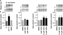
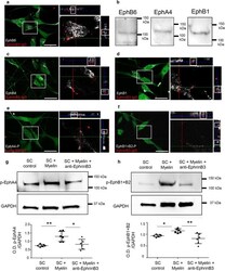
- EphA4 phosphorylation in primary spinal cord motor neurons (A and B) Representative Western blot images of pEphA4 and total EphA4 (after immuno-precipitation, IP). (B) graphs showing an average ratio of pEphA4 to total EphA4 in cultures of primary spinal cord motor neurons treated with DMSO, Fc (2.5 mug/mL), ephrinA1-Fc (eA1-Fc, 2.5 mug/mL), 123C4, 150E7, 150E8 (1 and 10 muM) and 150D4 (10 muM) for 15 min. Each panel has its own controls and error bars indicate SEM (each experiment was repeated 3-4 times). Statistical analysis was performed using one-way ANOVA followed by Bonferroni's post-hoc analysis (*p < 0.05, **p < 0.01, compared to DMSO).

- Submitted by
- Invitrogen Antibodies (provider)
- Main image

- Experimental details
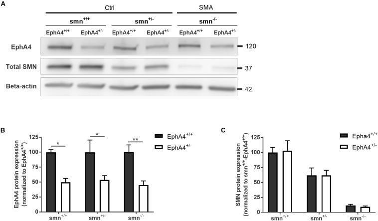
- FIGURE 1 EphA4 levels are reduced in the spinal cord of SMNDelta7 mice, without affecting SMN protein levels. Representative images (A) and quantifications (B,C) of a Western blot analysis of EphA4 and SMN levels in spinal cord lysates of Ctrl (smn +/+ and smn +/- ) and SMA (smn -/- ) pups are shown. Beta-actin protein levels were used as a loading control (two-way ANOVA with Sidak's multiple comparison test, n = 4-5 mice/group). * p
- Submitted by
- Invitrogen Antibodies (provider)
- Main image
- Experimental details
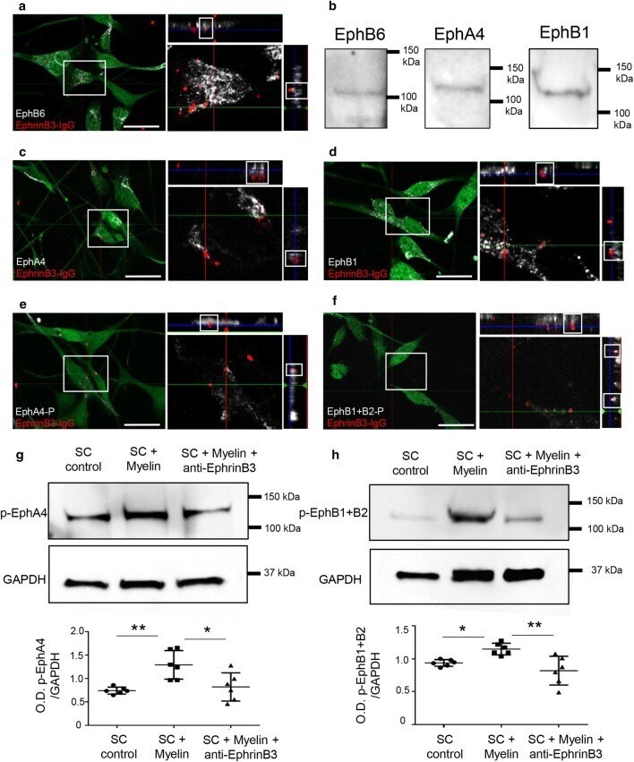
- Fig. 3 EphrinB3 binds and activates EphA4, EphB1 and EphB6 receptors in SC. a , c , d Orthogonal views of SC activation with clustered EphrinB3 shows that EphrinB3 binds EphB6 ( a ), EphA4 ( c ) and EphB1 ( d ) receptors on SC. b Western blot confirming the presence of these receptors in SC. e , f Bound EphrinB3 activation of EphA4 and EphB receptors viewed by immunodetection of phosphorylated forms. g , h Western blot illustrating that SC incubation with myelin increased the phosphorylation of EphA4 ( g ) and EphB1 + B2 ( h ) which was not the case when myelin was previously blocked by anti-EphrinB3 (O.D. p-EphB1 + B2/GAPDH: one-way ANOVA p = 0.003, F(2,15) = 8.57, followed by a Tukey's multiple comparison test) and O.D. of p-EphA4/GAPDH: one-way ANOVA p = 0.0036, F(2,15) = 8.44, followed by a Tukey's multiple comparison test). Data are expressed as ratio of the optical density (O.D.) of the bands (mean values +- SD) from three independent experiments, control ( n = 6), myelin ( n = 6), myelin + anti-EphrinB3 ( n = 6). Scale bar 20 um
- Submitted by
- Invitrogen Antibodies (provider)
- Main image

- Experimental details
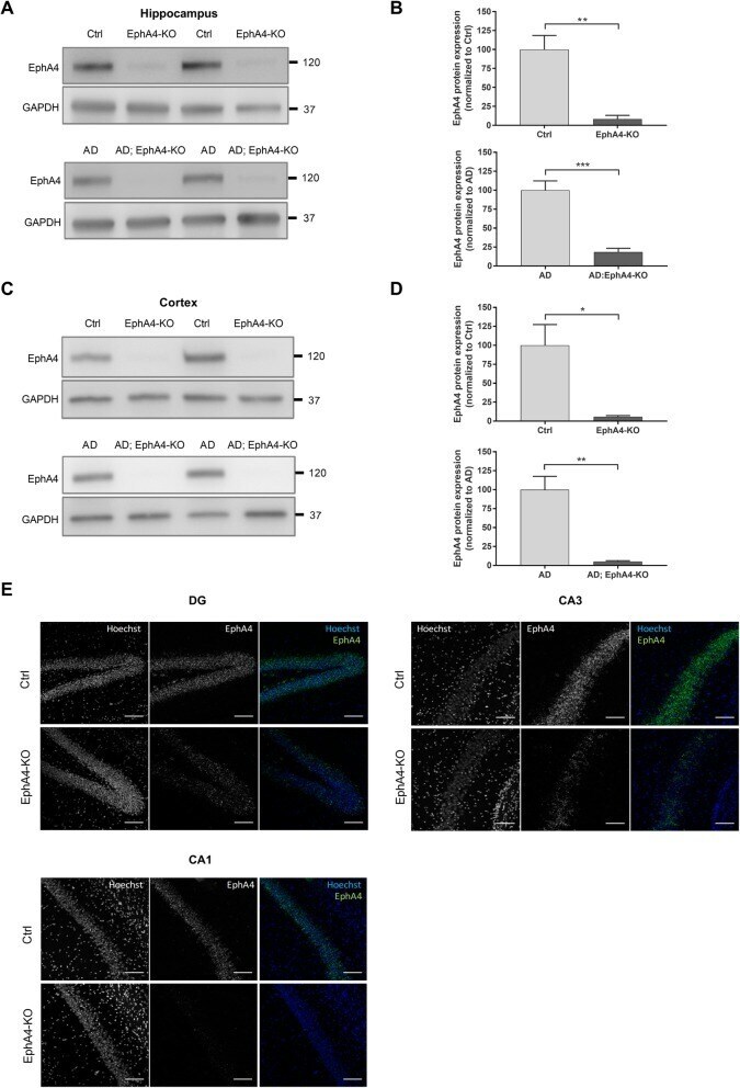
- Fig. 1 Reduction of EphA4 protein and mRNA levels in the hippocampus and cortex of APPPS1 mice. Representative images ( a ) and quantification ( b ) of a Western blot analysis of hippocampal EphA4 protein levels in Ctrl and EphA4-KO mice (upper blot) and AD and AD;EphA4-KO mice (lower blot) with GAPDH protein levels as a loading control (unpaired t test and Mann-Whitney test respectively, n = 4-8 mice/group). Representative images ( c ) and quantification ( d ) of a Western blot analysis of cortical EphA4 protein levels in Ctrl and EphA4-KO mice (upper blot) and AD and AD;EphA4-KO mice (lower blot). GAPDH protein levels were assessed to control for equal loading (unpaired t test, n = 4 mice/group). e Representative images of RNA scope with specific probes for EphA4 in the DG, CA3, and CA1 regions of the hippocampus of Ctrl and EphA4-KO mice. Hoechst was used to stain cell nuclei. * p
- Submitted by
- Invitrogen Antibodies (provider)
- Main image
- Experimental details
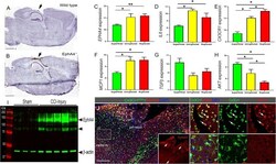
- Fig. 1 Neuroprotection in EphA4 -/- mice and human meta-analysis data of EPHA4. a , b Nissl staining of sagittal sections from WT or EphA4 global knockout mice at 14 days post-CCI injury compared to wild type. c - h Retrospective analysis of data archived as NIH GEO datasets from human patients following brain aneurysms. This study evaluated the gene expression on > 41,000 transcripts. Superficial, n = 10; unruptured, n = 5; ruptured, n = 8. i Western blot analysis for EphA4 protein expression in CCI-injured cortical tissue at 2 h compared to sham-injured cortices ( n = 3-5/group). j Representative confocal images of anti-EphA4 immuno-labeling (red; inset) at 24 h post-CCI injury in the ipsilateral cortex of CX3CR1 GFP/+ mice. CX3CR1-expressing microglia and/or infiltrating monocytes/macrophages show high expression of EphA4 in the peri-lesion cortex (J1) compared to adjacent cells in the medial parietal cortex (J2). Scale bar in j = 200 mum; scale bar in (J1) and (J2) = 20 mum. ANOVA with Bonferroni post hoc test. ** p < 0.01, *** p < 0.001 compared to superficial aneurysms

- Submitted by
- Invitrogen Antibodies (provider)
- Main image
- Experimental details
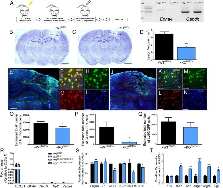
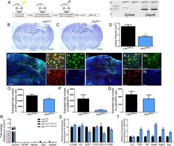
- Fig. 3 Bone marrow chimeric EphA4 -/- mice show decreased lesion volume and altered immune profile following CCI injury. a Bone marrow chimeric WT WTBMC and WT KOBMC mice were generated by irradiation and reconstitution with either wild-type or EphA4 knockout BMCs. PCR showing loss of transcript for Epha4 in the whole blood of WT KOBMC mice compared to WT WTBMC . b - d WT KOBMC mice display decreased lesion volumes compared to WT WTBMC at 3 days following CCI injury. e - i Max projection of z -stack confocal images of the injured cortex showing GFP+ BMCS and DAPI (blue) from WT and j - n KO immune cell infiltration. o Non-biased stereological quantification showed reduced total GFP+ numbers and p co-labeled CD68/GFP-positive cells in the ipsilateral cortex of WT KOBMC mice compared to WT WTBMC . q No difference was seen in the number of Ly6G/GFP+ neutrophils. r CD45-positive enriched cell isolation from the ipsilateral cortex at 3 days showed high purity of cx3cr1 mRNA expression compared with GFAP , Neun , Tie2 , and Vecad . s KO BMCs isolated from the injured cortex showed reduced pro-inflammatory CD68 , MCP1 , and Cxcl16 expression concomitant with t increased pro-resolving TGFbeta , Tie2 , Angpt1 , and Angpt2 . * p < 0.05, *** p < 0.001, **** p < 0.0001 compared to the corresponding WT BMCs. e and j scale bar = 500 mum; f - i and k - n scale bar = 50 mum