Explore
 Validate
 Learn
Learn Western blot
 ELISA
ELISAAntibody data
- Antibody Data
- Antigen structure
- References [13]
- Comments [0]
- Validations
- Western blot [1]
- Immunocytochemistry [1]
- Immunohistochemistry [2]
Submit
Validation data
Reference
Comment
Report error
- Product number
- 10384-1-AP - Provider product page

- Provider
- Proteintech Group
- Proper citation
- Proteintech Cat#10384-1-AP, RRID:AB_10638912
- Product name
- Cytokeratin 8 antibody
- Antibody type
- Polyclonal
- Description
- Cytokeratin 8 antibody (Cat. #10384-1-AP) is a rabbit polyclonal antibody that shows reactivity with human, mouse, rat and has been validated for the following applications: FC, IF, IHC, IP, WB, ELISA.
- Reactivity
- Human, Mouse, Rat
- Host
- Rabbit
- Conjugate
- Unconjugated
- Isotype
- IgG
- Vial size
- 20ul, 150ul
Submitted references Unveiling the Role of Vitamin D/VDR in Promoting Endometrial Decidualization.
Crip2 affects vascular development by fine-tuning endothelial cell aggregation and proliferation.
Effect of defective H2B ubiquitination on the malignancy and repair of DNA double breaks and its significance in lung adenocarcinoma.
Luminal hormone-responsive cells tune the regenerative remodeling of mammary glands in large mammals.
An integrated approach identifies the molecular underpinnings of murine anterior visceral endoderm migration.
D-tagatose protects against oleic acid-induced acute respiratory distress syndrome in rats by activating PTEN/PI3K/AKT pathway.
COVID-19 lung disease shares driver AT2 cytopathic features with Idiopathic pulmonary fibrosis.
Comparative study of the external auditory canal in humans and large mammals.
COVID-19 lung disease shares driver AT2 cytopathic features with Idiopathic pulmonary fibrosis.
ESRRB Facilitates the Conversion of Trophoblast-Like Stem Cells From Induced Pluripotent Stem Cells by Directly Regulating CDX2.
The helix-loop-helix transcriptional regulator Id4 is required for terminal differentiation of luminal epithelial cells in the prostate.
SNAP29 mediates the assembly of histidine-induced CTP synthase filaments in proximity to the cytokeratin network.
Alternative promotion and suppression of metastasis by JNK2 governed by its phosphorylation.
Guo J, Tian X, Liu H, Lu Q, Cao G
International journal of endocrinology 2026;2026:1712178
International journal of endocrinology 2026;2026:1712178
Crip2 affects vascular development by fine-tuning endothelial cell aggregation and proliferation.
Yang S, Zhang X, Li X, Li H
Cellular and molecular life sciences : CMLS 2025 Mar 13;82(1):110
Cellular and molecular life sciences : CMLS 2025 Mar 13;82(1):110
Effect of defective H2B ubiquitination on the malignancy and repair of DNA double breaks and its significance in lung adenocarcinoma.
An Z, Yao N, Su W, Zhang W, Yang J, Guo Q, Zhang C, Tian P, Wan N, Wu C, Huang X, Song G, Jia J, Ren L
Scientific reports 2025 Jul 2;15(1):22576
Scientific reports 2025 Jul 2;15(1):22576
Luminal hormone-responsive cells tune the regenerative remodeling of mammary glands in large mammals.
Li Y, Zhang L, Luo T, Zhang W, Wang T, Liu F, Lin S, Luo J, Liu J, Peng J, Wang C, Wang W, Shi H
Cell discovery 2025 Dec 30;11(1):105
Cell discovery 2025 Dec 30;11(1):105
An integrated approach identifies the molecular underpinnings of murine anterior visceral endoderm migration.
Thowfeequ S, Fiorentino J, Hu D, Solovey M, Ruane S, Whitehead M, Zhou F, Godwin J, Mateo-Otero Y, Vanhaesebroeck B, Scialdone A, Srinivas S
Developmental cell 2024 Sep 9;59(17):2347-2363.e9
Developmental cell 2024 Sep 9;59(17):2347-2363.e9
D-tagatose protects against oleic acid-induced acute respiratory distress syndrome in rats by activating PTEN/PI3K/AKT pathway.
Huang J, Wang B, Tao S, Hu Y, Wang N, Zhang Q, Wang C, Chen C, Gao B, Cheng X, Li Y
Frontiers in immunology 2022;13:928312
Frontiers in immunology 2022;13:928312
COVID-19 lung disease shares driver AT2 cytopathic features with Idiopathic pulmonary fibrosis.
Sinha S, Castillo V, Espinoza CR, Tindle C, Fonseca AG, Dan JM, Katkar GD, Das S, Sahoo D, Ghosh P
bioRxiv : the preprint server for biology 2022 Jul 18;
bioRxiv : the preprint server for biology 2022 Jul 18;
Comparative study of the external auditory canal in humans and large mammals.
Wang J, He A, Yin D, Zhu Y, Zhou G, Zhang T
Anatomical record (Hoboken, N.J. : 2007) 2022 Feb;305(2):436-445
Anatomical record (Hoboken, N.J. : 2007) 2022 Feb;305(2):436-445
COVID-19 lung disease shares driver AT2 cytopathic features with Idiopathic pulmonary fibrosis.
Sinha S, Castillo V, Espinoza CR, Tindle C, Fonseca AG, Dan JM, Katkar GD, Das S, Sahoo D, Ghosh P
EBioMedicine 2022 Aug;82:104185
EBioMedicine 2022 Aug;82:104185
ESRRB Facilitates the Conversion of Trophoblast-Like Stem Cells From Induced Pluripotent Stem Cells by Directly Regulating CDX2.
Yu S, Zhang R, Shen Q, Zhu Z, Zhang J, Wu X, Zhao W, Li N, Yang F, Wei H, Hua J
Frontiers in cell and developmental biology 2021;9:712224
Frontiers in cell and developmental biology 2021;9:712224
The helix-loop-helix transcriptional regulator Id4 is required for terminal differentiation of luminal epithelial cells in the prostate.
Hewa Bostanthirige D, Komaragiri SK, Joshi JB, Alzahrani M, Saini I, Jain S, Bowen NJ, Havrda MC, Chaudhary J
Oncoscience 2021;8:14-30
Oncoscience 2021;8:14-30
SNAP29 mediates the assembly of histidine-induced CTP synthase filaments in proximity to the cytokeratin network.
Chakraborty A, Lin WC, Lin YT, Huang KJ, Wang PY, Chang IY, Wang HI, Ma KT, Wang CY, Huang XR, Lee YH, Chen BC, Hsieh YJ, Chien KY, Lin TY, Liu JL, Sung LY, Yu JS, Chang YS, Pai LM
Journal of cell science 2020 May 11;133(9)
Journal of cell science 2020 May 11;133(9)
Alternative promotion and suppression of metastasis by JNK2 governed by its phosphorylation.
Hu S, Dong X, Gao W, Stupack D, Liu Y, Xiang R, Li N
Oncotarget 2017 Aug 22;8(34):56569-56581
Oncotarget 2017 Aug 22;8(34):56569-56581
No comments: Submit comment
Supportive validation
- Submitted by
- Proteintech Group (provider)
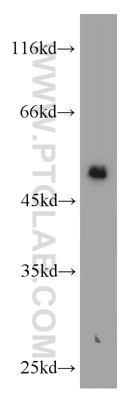
- Main image

- Experimental details
- MCF7 cells were subjected to SDS PAGE followed by western blot with 10384-1-AP(KRT8 antibody) at dilution of 1:1500
- Sample type
- cell line
Supportive validation
- Submitted by
- Proteintech Group (provider)
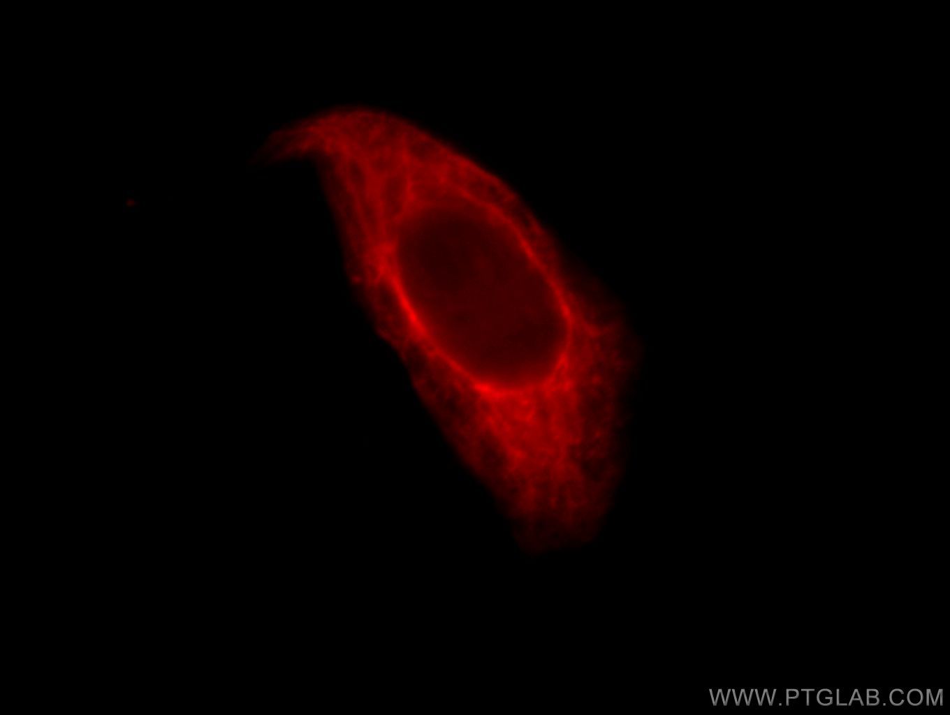
- Main image
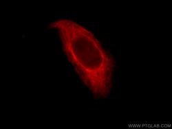
- Experimental details
- Immunofluorescent analysis of HepG2 cells, using KRT8 antibody 10384-1-AP at 1:25 dilution and Rhodamine-labeled goat anti-rabbit IgG (red).
- Sample type
- cell line
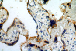
Supportive validation
- Submitted by
- Proteintech Group (provider)
- Main image
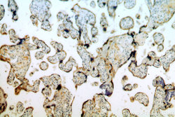
- Experimental details
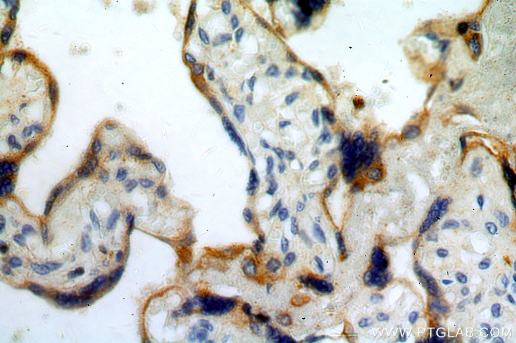
- Immunohistochemical of paraffin-embedded human placenta using 10384-1-AP(KRT8 antibody) at dilution of 1:100 (under 10x lens)
- Sample type
- tissue
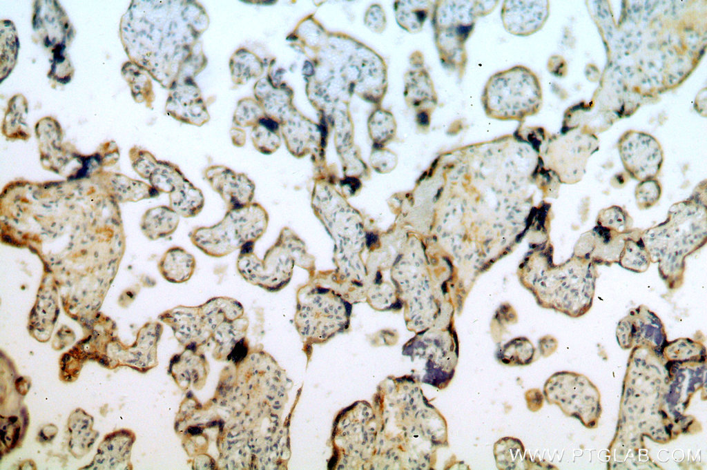
- Submitted by
- Proteintech Group (provider)
- Main image
- Experimental details
- Immunohistochemical of paraffin-embedded human placenta using 10384-1-AP(KRT8 antibody) at dilution of 1:100 (under 40x lens)
- Sample type
- tissue