Explore
 Validate
 Learn
Learn Western blot
Antibody data
- Antibody Data
- Antigen structure
- References [0]
- Comments [0]
- Validations
- Western blot [2]
- Immunocytochemistry [2]
- Immunohistochemistry [6]
- Other assay [5]
Submit
Validation data
Reference
Comment
Report error
- Product number
- MA5-13156 - Provider product page

- Provider
- Invitrogen Antibodies
- Product name
- Cytokeratin Pan Type I/II Antibody Cocktail
- Antibody type
- Monoclonal
- Antigen
- Other
- Description
- MA5-13156 targets Cytokeratin Pan in WB, IF and IHC (P) applications and shows reactivity with Bovine, Chicken, Human, mouse, Non-human primate, Rabbit, and Rat samples. This antibody is not suitable for PC12 cells in IF or WB applications. The MA5-13156 immunogen is human epidermal keratin. This antibody detects the acidic and basic (Type I and II) cytokeratins: Cytokeratin 1, Cytokeratin 2, Cytokeratin 3, Cytokeratin 4, Cytokeratin 5, Cytokeratin 6, Cytokeratin 7, Cytokeratin 8, Cytokeratin 10, Cytokeratin 14, Cytokeratin 15, Cytokeratin 16 and Cytokeratin 19.
- Reactivity
- Human, Mouse
- Host
- Mouse
- Isotype
- IgG
- Antibody clone number
- AE1/AE3
- Vial size
- 500 µL
- Concentration
- 0.2 mg/mL
- Storage
- 4° C
No comments: Submit comment
Supportive validation
- Submitted by
- Invitrogen Antibodies (provider)
- Main image
- Experimental details
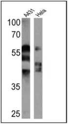
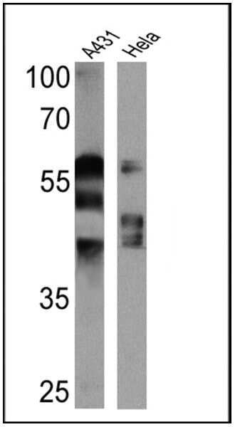
- Western blot analysis of Cytokeratin Pan was performed by loading 25 µg of A431 (Lane 1) and Hela (Lane 2) cell lysates onto an SDS polyacrylamide gel. Proteins were transferred to a PVDF membrane and blocked at 4ºC overnight. The membrane was probed with a Cytokeratin Pan monoclonal antibody (Product # MA5-13156) at a dilution of 1:2000 overnight at 4°C, washed in TBST, and probed with an HRP-conjugated secondary antibody for 1 hr at room temperature in the dark. Chemiluminescent detection was performed using Pierce ECL Plus Western Blotting Substrate (Product # 32132). Results show a band at approx. 40-67 kDa.
- Submitted by
- Invitrogen Antibodies (provider)
- Main image
- Experimental details
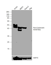
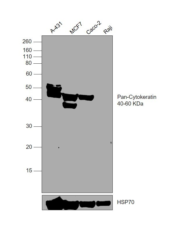
- Western blot was performed using Anti-Cytokeratin Pan Type I/II Antibody Cocktail (Product # MA5-13156) and a 40-60 kDa band corresponding to Cytokeratin Pan Type I/II was observed across all tested cell lines, except Raji. Whole cell extracts (30 µg lysate) of A-431 (Lane 1), MCF7 (Lane 2), Caco-2 (Lane 3), Raji (Lane 4) were electrophoresed using NuPAGE™ 10% Bis-Tris Protein Gel (Product # NP0301BOX). Resolved proteins were then transferred onto a nitrocellulose membrane (Product # IB23002) by iBlot® 2 Dry Blotting System (Product # IB21001). The blot was probed with the primary antibody (1:1000 dilution) and detected by chemiluminescence with Goat anti-Mouse IgG (H+L) Superclonal™ Recombinant Secondary Antibody, HRP (Product # A28177,1:10000 dilution) using the iBright™ FL1500 Imaging System (Product # A44115). Chemiluminescent detection was performed using SuperSignal™ West Pico PLUS Chemiluminescent Substrate (Product # 34580).
Supportive validation
- Submitted by
- Invitrogen Antibodies (provider)
- Main image
- Experimental details
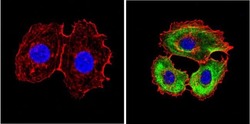
- Immunofluorescent analysis of Cytokeratin Pan (green) showing positive staining in the cytoplasm of MCF-7 cells (right) compared with a negative control in the absence of primary antibody (left). Formalin-fixed cells were permeabilized with 0.1% Triton X-100 in TBS for 5-10 minutes, blocked with 3% BSA-PBS for 30 minutes at room temperature and probed with a Cytokeratin Pan monoclonal antibody (Product # MA5-13156) in 3% BSA-PBS at a dilution of 1:100 and incubated overnight at 4 ºC in a humidified chamber. Cells were washed with PBST and incubated with a DyLight 488-conjugated goat-anti-mouse IgG (H+L) secondary antibody in PBS at room temperature in the dark. F-actin (red) was stained with a fluorescent red phalloidin and nuclei (blue) were stained with DAPI for 5-10 minutes in the dark. Images were taken at a magnification of 60x.
- Submitted by
- Invitrogen Antibodies (provider)
- Main image

- Experimental details
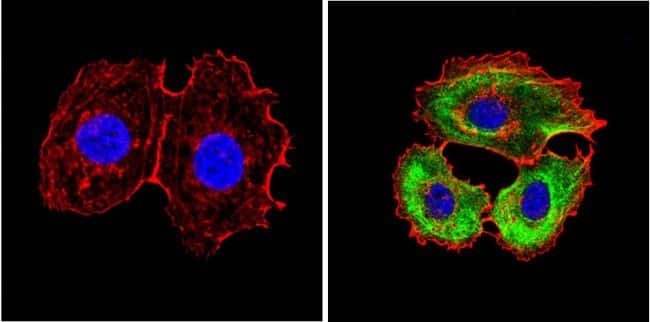
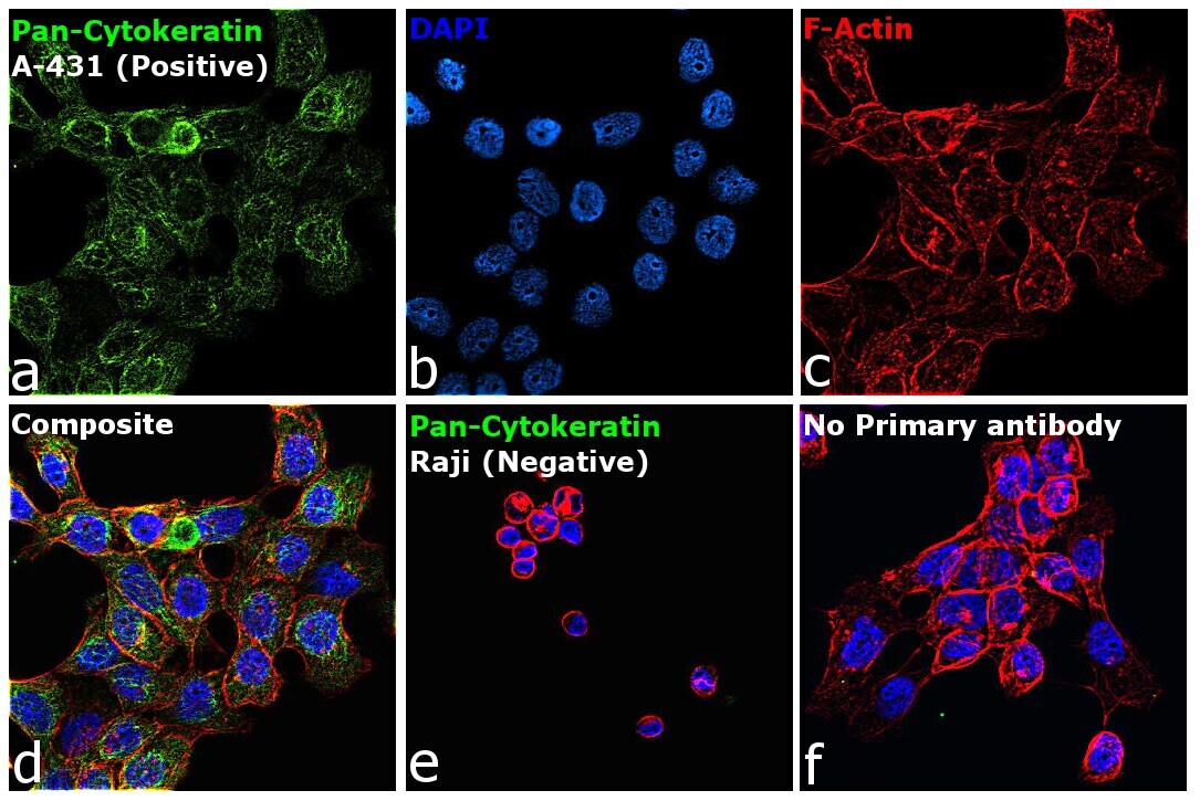
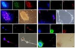
- Immunofluorescence analysis of Cytokeratin Pan Type I/II was performed using 70% confluent log phase A-431 cells. The cells were fixed and permeabilized with ice-cold acetone at 4°C for 5 minutes, and blocked with 2% BSA for 1 hour at room temperature. The cells were labeled with Cytokeratin Pan Type I/II Antibody Cocktail (Product # MA5-13156, 5 µg/mL) in 0.1% BSA, incubated at 4 degree celsius overnight and then labeled with Donkey anti-Mouse IgG (H+L) Highly Cross-Adsorbed Secondary Antibody, Alexa Fluor Plus 488 (Product # A32766, 1:2000 dilution), for 45 minutes at room temperature (Panel a: Green). Nuclei (Panel b: Blue) were stained with ProLong™ Diamond Antifade Mountant with DAPI (Product # P36962). F-actin (Panel c: Red) was stained with Rhodamine Phalloidin (Product # R415, 1:300 dilution). Panel d represents the merged image showing cytoskeletal localization. Panel e represents Raji cells showing no expression of cytokeratin. Panel f represents control cells with no primary antibody to assess background. The images were captured at 60X magnification.
Supportive validation
- Submitted by
- Invitrogen Antibodies (provider)
- Main image

- Experimental details
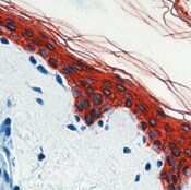
- Formalin-fixed, paraffin-embedded human skin stained with Keratin, Pan antibody using peroxidase-conjugate and AEC chromogen. Note cytoplasmic staining of epithelial cells.
- Submitted by
- Invitrogen Antibodies (provider)
- Main image

- Experimental details
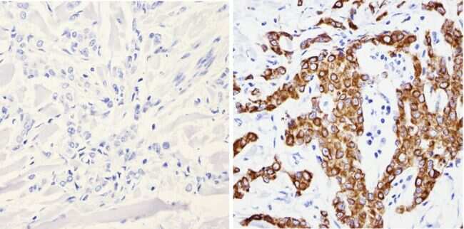
- Immunohistochemistry analysis of Cytokeratin Pan showing positive staining in the cytoplasm of paraffin-treated Human breast carcinoma (right) compared with a negative control in the absence of primary antibody (left). To expose target proteins, antigen retrieval method was performed using 10mM sodium citrate (pH 6.0) microwaved for 8-15 min. Following antigen retrieval, tissues were blocked in 3% H2O2-methanol for 15 min at room temperature, washed with ddH2O and PBS, and then probed with a Cytokeratin Pan monoclonal antibody (Product # MA5-13156) diluted by 3% BSA-PBS at a dilution of 1:200 overnight at 4°C in a humidified chamber. Tissues were washed extensively PBST and detection was performed using an HRP-conjugated secondary antibody followed by colorimetric detection using a DAB kit. Tissues were counterstained with hematoxylin and dehydrated with ethanol and xylene to prep for mounting.
- Submitted by
- Invitrogen Antibodies (provider)
- Main image
- Experimental details
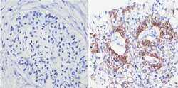
- Immunohistochemistry analysis of Cytokeratin Pan showing positive staining in the cytoplasm of paraffin-treated Human lung squamous carcinoma (right) compared with a negative control in the absence of primary antibody (left). To expose target proteins, antigen retrieval method was performed using 10mM sodium citrate (pH 6.0) microwaved for 8-15 min. Following antigen retrieval, tissues were blocked in 3% H2O2-methanol for 15 min at room temperature, washed with ddH2O and PBS, and then probed with a Cytokeratin Pan monoclonal antibody (Product # MA5-13156) diluted by 3% BSA-PBS at a dilution of 1:200 overnight at 4°C in a humidified chamber. Tissues were washed extensively PBST and detection was performed using an HRP-conjugated secondary antibody followed by colorimetric detection using a DAB kit. Tissues were counterstained with hematoxylin and dehydrated with ethanol and xylene to prep for mounting.
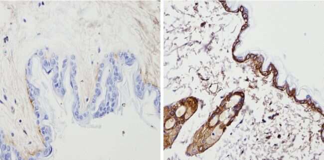
- Submitted by
- Invitrogen Antibodies (provider)
- Main image

- Experimental details
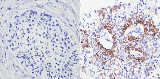
- Immunohistochemistry analysis of Cytokeratin Pan showing positive staining in the cytoplasm of paraffin-treated Mouse skin tissue (right) compared with a negative control in the absence of primary antibody (left). To expose target proteins, antigen retrieval method was performed using 10mM sodium citrate (pH 6.0) microwaved for 8-15 min. Following antigen retrieval, tissues were blocked in 3% H2O2-methanol for 15 min at room temperature, washed with ddH2O and PBS, and then probed with a Cytokeratin Pan monoclonal antibody (Product # MA5-13156) diluted by 3% BSA-PBS at a dilution of 1:200 overnight at 4°C in a humidified chamber. Tissues were washed extensively PBST and detection was performed using an HRP-conjugated secondary antibody followed by colorimetric detection using a DAB kit. Tissues were counterstained with hematoxylin and dehydrated with ethanol and xylene to prep for mounting.
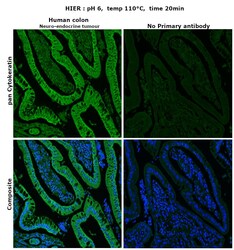
- Submitted by
- Invitrogen Antibodies (provider)
- Main image
- Experimental details
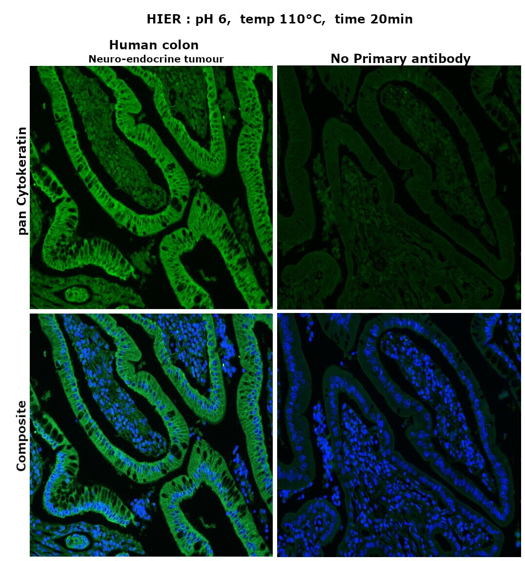
- Immunohistochemical analysis of pan Cytokeratin was performed using formalin-fixed paraffin-embedded human colon (neuro-endocrine tumor) tissue sections. To expose the target protein, heat-induced epitope retrieval (HIER) was performed on de-paraffinized sections using eBioscience™ IHC Antigen Retrieval Solution - Low pH (10X) (Product # 00-4955-58) diluted to 1X solution in water in a decloaking chamber at 110 degree Celsius for 20 minutes. Following antigen retrieval, the sections were blocked with 2% normal goat serum in 1X PBS for 45 minutes at room temperature and then probed with or without Cytokeratin Pan Type I/II Antibody Cocktail (Product # MA5-13156) at 2 µg/mL in 0.1% normal goat serum overnight at 4 degree Celsius in a humidified chamber. Detection was performed using Goat anti-Mouse IgG (H+L) Highly Cross-Adsorbed Secondary Antibody, Alexa Fluor Plus 488 (Product # A32723) at a dilution of 1:2000 in 0.1% normal goat serum for 45 minutes at room temperature. Nuclei were stained with DAPI (Product # D1306) and the sections were mounted using ProLong™ Glass Antifade Mountant (Product # P36984). The images were captured on EVOS™ M7000 Imaging System (Product # AMF7000) at 20X magnification.
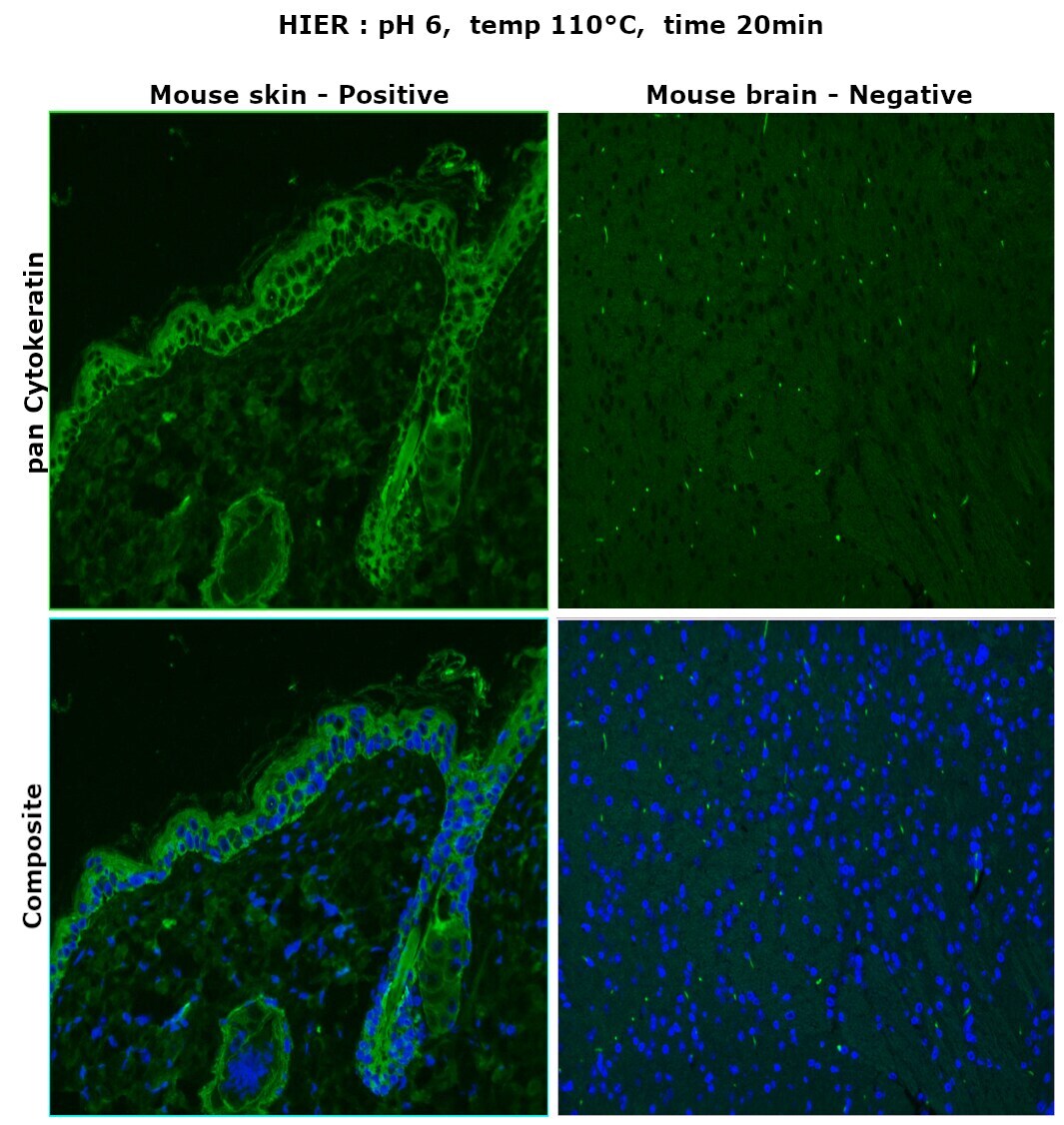
- Submitted by
- Invitrogen Antibodies (provider)
- Main image

- Experimental details
- Immunohistochemical analysis of pan Cytokeratin was performed using formalin-fixed paraffin-embedded mouse skin and mouse brain tissue sections. To expose the target protein, heat-induced epitope retrieval (HIER) was performed on de-paraffinized sections using eBioscience™ IHC Antigen Retrieval Solution - Low pH (10X) (Product # 00-4955-58) diluted to 1X solution in water in a decloaking chamber at 110 degree Celsius for 20 minutes. Following antigen retrieval, the sections were blocked with 2% normal goat serum in 1X PBS for 45 minutes at room temperature and then probed with Cytokeratin Pan Type I/II Antibody Cocktail (Product # MA5-13156) at 4 µg/mL in 0.1% normal goat serum overnight at 4 degree Celsius in a humidified chamber. Detection was performed using Goat anti-Mouse IgG (H+L) Highly Cross-Adsorbed Secondary Antibody, Alexa Fluor Plus 488 (Product # A32723) at a dilution of 1:2000 in 0.1% normal goat serum for 45 minutes at room temperature. Nuclei were stained with DAPI (Product # D1306) and the sections were mounted using ProLong™ Glass Antifade Mountant (Product # P36984). The images were captured on EVOS™ M7000 Imaging System (Product # AMF7000) at 20X magnification.
Supportive validation
- Submitted by
- Invitrogen Antibodies (provider)
- Main image
- Experimental details
- NULL

- Submitted by
- Invitrogen Antibodies (provider)
- Main image
- Experimental details
- NULL

- Submitted by
- Invitrogen Antibodies (provider)
- Main image
- Experimental details
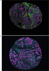
- FIGURE 1 Representative immunofluorescence images. (A) Representative image of a TMA core with heterogeneous infiltration of leukocytes in panel 1; red denoting CD68, yellow denoting CD56, green denoting CD3, pink denoting NKp46, cyan denoting CD163 and magenta denoting cytokeratin. (B) Representative image of a TMA core with heterogeneous infiltration of leukocytes in panel 2; green denoting CD1a, red denoting CD208, yellow denoting CD15, pink denoting CD123, cyan denoting CD68 and magenta denoting cytokeratin.

- Submitted by
- Invitrogen Antibodies (provider)
- Main image
- Experimental details
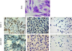
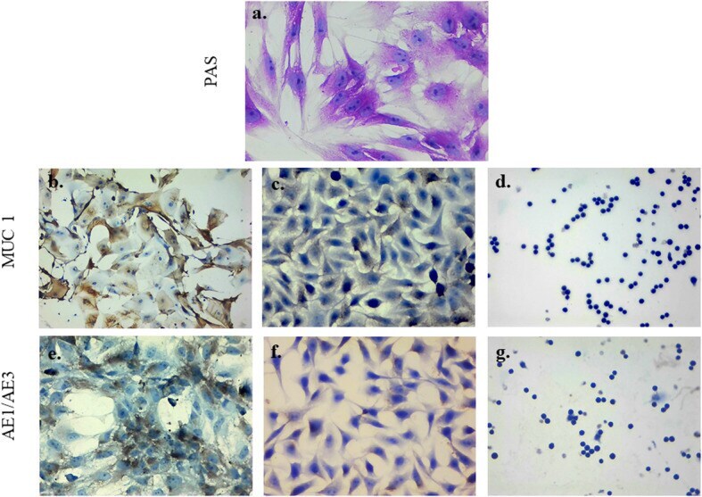
- Fig. 5 Mucin production and epithelial phenotype in GECs. a. GEC were stained with PAS (reddish-purple staining indicates the presence of neutral mucins), b. MUC1 on GECs shows homogeneous cytoplasmic staining mainly located in the cell membrane, c. AGS cell lines (mucin positive control) and d. U-937 cell lines (negative control) e . Pan-cytokeratin markers AE1/AE3 on GECs: a positive fibrillar staining pattern distributed in the cytoplasm is observed, f. HeLa cell lines (positive control) and, g. U-937 cell lines (negative control). Images were visualized using conventional light microscopy. Images were taken at 20x magnification
- Submitted by
- Invitrogen Antibodies (provider)
- Main image
- Experimental details
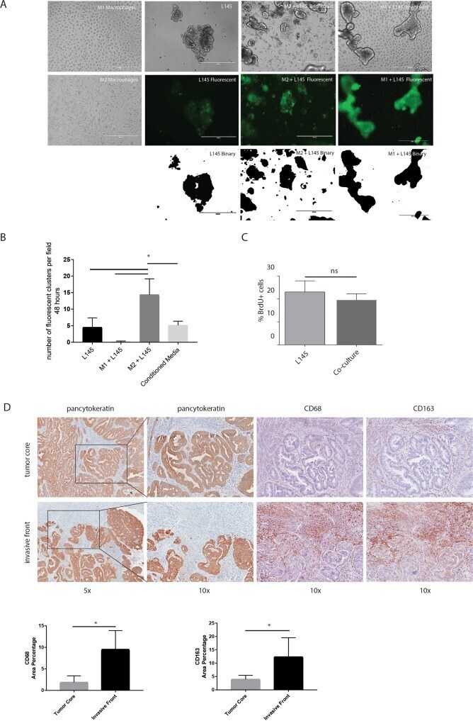
- Figure 3 M2 macrophages cause tumor cell budding from the colonosphere bulk (A) Representative images of bright field microscopy of the mono-cultures and co-cultures are shown in the upper panel. Green fluorescent images to identify tumor cells are shown in the middle panel, binarized images of the fluorescence are shown in the lower panel, 10x magnification on EVOS microscope; scale bars indicate 400 mum. (B) Adherence assay showing the number of small cell clusters (