Explore
 Validate
 Learn
Learn Western blot
 Flow cytometry
Antibody data
- Antibody Data
- Antigen structure
- References [2]
- Comments [0]
- Validations
- Flow cytometry [1]
Submit
Validation data
Reference
Comment
Report error
- Product number
- NB120-9287 - Provider product page

- Provider
- Novus Biologicals
- Proper citation
- Novus Cat#NB120-9287, RRID:AB_2134857
- Product name
- Mouse Monoclonal Cytokeratin 8 Antibody
- Antibody type
- Monoclonal
- Description
- Protein A or G purified. Recognizes most of the basic (Type II) keratins, HMW cytokeratins. It is broadly reactive and stains positively almost all epithelia and their neoplasms. AE3 reacts with all layers of epidermis (basal layer and above). The antibody is an excellent marker for distinguishing carcinomas from non-epithelial tumours. It has been used for studying the immunolocalisation of epidermal keratins, tissue distribution of keratins, various diseases of epidermis, oral mucosa and other epithelia and other epithelia development.
- Reactivity
- Human, Mouse, Rat, Bovine, Chicken/Avian, Rabbit, Simian
- Host
- Mouse
- Isotype
- IgG
- Vial size
- 0.1 mg
- Concentration
- 1.0 mg/ml
- Storage
- Store at 4C short term. Aliquot and store at -20C long term. Avoid freeze-thaw cycles.
Submitted references The common parasite Toxoplasma gondii induces prostatic inflammation and microglandular hyperplasia in a mouse model.
Chemoprevention activity of 25-hydroxyvitamin D in the MMTV-PyMT mouse model of breast cancer.
Colinot DL, Garbuz T, Bosland MC, Wang L, Rice SE, Sullivan WJ Jr, Arrizabalaga G, Jerde TJ
The Prostate 2017 Jul;77(10):1066-1075
The Prostate 2017 Jul;77(10):1066-1075
Chemoprevention activity of 25-hydroxyvitamin D in the MMTV-PyMT mouse model of breast cancer.
Rossdeutscher L, Li J, Luco AL, Fadhil I, Ochietti B, Camirand A, Huang DC, Reinhardt TA, Muller W, Kremer R
Cancer prevention research (Philadelphia, Pa.) 2015 Feb;8(2):120-8
Cancer prevention research (Philadelphia, Pa.) 2015 Feb;8(2):120-8
No comments: Submit comment
Supportive validation
- Submitted by
- Novus Biologicals (provider)
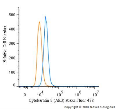
- Main image
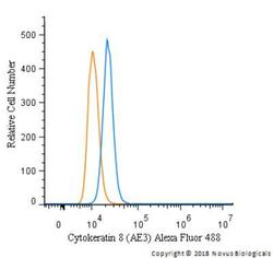
- Experimental details
- Flow Cytometry: Cytokeratin 8 Antibody (AE3) [NB120-9287] - An intracellular stain was performed on A431 cells with Cytokeratin 8 Antibody [AE3] NB120-9287AF488 (blue) and a matched isotype control (orange). Cells were fixed with 4% PFA and then permeabilized with 0.1% saponin. Cells were incubated in an antibody dilution of 5 ug/mL for 30 minutes at room temperature. Both antibodies were conjugated to Alexa Fluor 488.