Explore
 Validate
 Learn
Learn Western blot
Antibody data
- Antibody Data
- Antigen structure
- References [1]
- Comments [0]
- Validations
- Western blot [1]
- Immunocytochemistry [1]
Submit
Validation data
Reference
Comment
Report error
- Product number
- 702176 - Provider product page

- Provider
- Invitrogen Antibodies
- Product name
- Phospho-Cytokeratin 8 (Ser432) Recombinant Rabbit Monoclonal Antibody (21H19L3)
- Antibody type
- Monoclonal
- Antigen
- Synthetic peptide
- Description
- This antibody is predicted to react with Monkey, Horse, Mouse
- Antibody clone number
- 21H19L3
- Concentration
- 0.5 mg/mL
Submitted references Expression of keratins 8, 18, and 19 in epithelia of atrophic oral lichen planus.
Schreurs O, Karatsaidis A, Balta MG, Grung B, Hals EKB, Schenck K
European journal of oral sciences 2020 Feb;128(1):7-17
European journal of oral sciences 2020 Feb;128(1):7-17
No comments: Submit comment
Supportive validation
- Submitted by
- Invitrogen Antibodies (provider)
- Main image
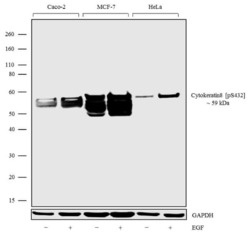
- Experimental details
- Western blot analysis was performed on Whole cell extracts (30 µg lysate) of Caco-2 (Lane 1), Caco-2 treated with EGF (100 ng/mL for 20 min) (Lane 2), MCF7 (Lane 3), MCF7 treated with EGF (100 ng/mL for 20 min) (Lane 4), HeLa (Lane 5) and HeLa treated with EGF (100 ng/mL for 20 min) (Lane 6). The blots were probed with Anti-Phospho-Cytokeratin 8 (Ser432) Rabbit Monoclonal Antibody (Product # 702176, 1-2 µg/mL) and detected by chemiluminescence using Goat anti-Rabbit IgG (H+L) Superclonal Secondary Antibody, HRP conjugate (Product # A27036, 0.4 µg/mL, 1:2500 dilution). A 59 kDa band corresponding to Phospho-Cytokeratin 8 (Ser432) was observed across the cell lines tested. Known quantity of protein samples were electrophoresed using Novex® NuPAGE® 4-12% Bis-Tris gel (Product # NP0321BOX), XCell SureLock Electrophoresis System (Product # EI0002) and Novex® Sharp Pre-Stained Protein Standard (Product # LC5800). Resolved proteins were then transferred onto a nitrocellulose membrane with iBlot® Dry Blotting System (Product # IB21001). The membrane was probed with the relevant primary and secondary Antibody following blocking with 5% skimmed milk. Chemiluminescent detection was performed using Pierce™ ECL Western Blotting Substrate (Product # 32106).

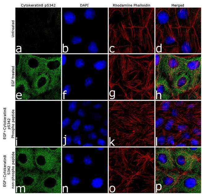
Supportive validation
- Submitted by
- Invitrogen Antibodies (provider)
- Main image

- Experimental details
- For immunofluorescence analysis CACO-2 cells were fixed and permeabilized for detection of endogenous Phospho-Cytokeratin 8 (Ser432) using Anti-Phospho-Cytokeratin 8 (Ser432) Recombinant Rabbit Monoclonal Antibody (Product # 702176, 2 µg/mL) and labeled with Goat anti-Rabbit IgG (H+L) Superclonal Secondary Antibody, Alexa Fluor® 488 conjugate (Product # A27034, 1:2000). Nuclei (blue) were stained using SlowFade® Gold Antifade Mountant with DAPI (Product # S36938), and Rhodamine Phalloidin (Product # R415, 1:300) was used for cytoskeletal F-actin (red) staining. Detection and localization of Phospho-Cytokeratin 8 (Ser432) (green) in the cytoskeleton can be clearly observed in cells treated with EGF (100 ng/mL, 20 min) as compared to untreated cells. Antibody specificity was demonstrated by competition with the Cytokeratin 8 (Ser432) phospho peptide, which results in loss of signal. No competition was observed with the non-phospho peptide. The images were captured at 60X magnification.