Explore
 Validate
 Learn
Learn Western blot
Antibody data
- Antibody Data
- Antigen structure
- References [1]
- Comments [0]
- Validations
- Western blot [4]
- Immunocytochemistry [1]
- Immunohistochemistry [1]
Submit
Validation data
Reference
Comment
Report error
- Product number
- GTX110311 - Provider product page

- Provider
- GeneTex
- Proper citation
- GeneTex Cat#GTX110311, RRID:AB_11165171
- Product name
- Cytokeratin 8 antibody
- Antibody type
- Polyclonal
- Reactivity
- Human, Mouse
- Host
- Rabbit
Submitted references Cdc6 cooperates with c-Myc to promote genome instability and epithelial to mesenchymal transition EMT in zebrafish.
Chen CH, Lin DS, Cheng CW, Lin CJ, Lo YK, Yen CC, Lee AY, Hsiao CD
Oncotarget 2014 Aug 15;5(15):6300-11
Oncotarget 2014 Aug 15;5(15):6300-11
No comments: Submit comment
Supportive validation
- Submitted by
- GeneTex (provider)
- Main image
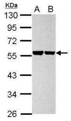
- Experimental details
- Sample (30 ?g of whole cell lysate) A: HeLa B: HepG2 10% SDS PAGE GTX110311 diluted at 1:10000 The HRP-conjugated anti-rabbit IgG antibody (GTX213110-01) was used to detect the primary antibody.

- Submitted by
- GeneTex (provider)
- Main image
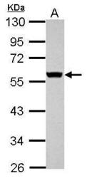
- Experimental details
- Sample (30 ?g of whole cell lysate) A: JC 10% SDS PAGE GTX110311 diluted at 1:10000 The HRP-conjugated anti-rabbit IgG antibody (GTX213110-01) was used to detect the primary antibody.
- Submitted by
- GeneTex (provider)
- Main image
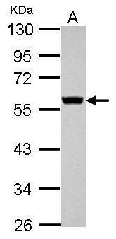
- Experimental details
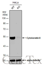
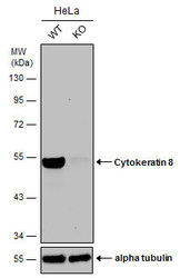
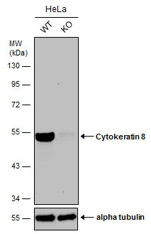
- Wild-type (WT) and Cytokeratin 8 knockout (KO) HeLa cell extracts (30 ?g) were separated by 10% SDS-PAGE, and the membrane was blotted with Cytokeratin 8 antibody (GTX110311) diluted at 1:20000. The HRP-conjugated anti-rabbit IgG antibody (GTX213110-01) was used to detect the primary antibody.
- Submitted by
- GeneTex (provider)
- Main image
- Experimental details
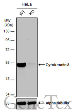
- Wild-type (WT) and Cytokeratin 8 knockout (KO) HeLa cell extracts (30 ?g) were separated by 10% SDS-PAGE, and the membrane was blotted with Cytokeratin 8 antibody (GTX110311) diluted at 1:20000. The HRP-conjugated anti-rabbit IgG antibody (GTX213110-01) was used to detect the primary antibody.
Supportive validation
- Submitted by
- GeneTex (provider)
- Main image
- Experimental details
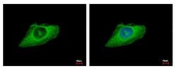
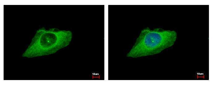
- Cytokeratin 8 antibody detects KRT8 protein at cytoplasm by immunofluorescent analysis. Sample: HeLa cells were fixed in ice-cold MeOH for 5 min.Green: KRT8 protein stained by Cytokeratin 8 antibody (GTX110311) diluted at 1:500.Blue: Hoechst 33343 staining.
Supportive validation
- Submitted by
- GeneTex (provider)
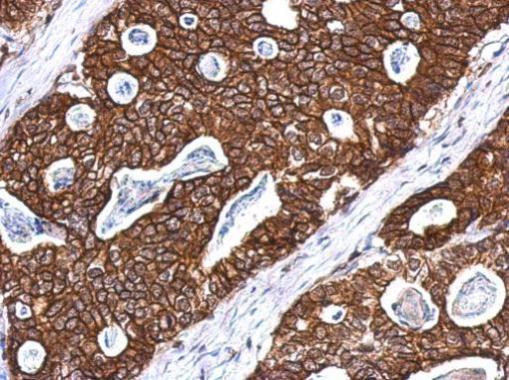
- Main image
- Experimental details
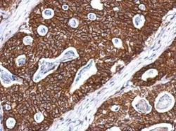
- Immunohistochemical analysis of paraffin-embedded Gastric ca, using Cytokeratin 8(GTX110311) antibody at 1:500 dilution.