Explore
 Validate
 Learn
Learn Western blot
Antibody data
- Antibody Data
- Antigen structure
- References [1]
- Comments [0]
- Validations
- Western blot [3]
- Immunocytochemistry [1]
- Immunohistochemistry [1]
Submit
Validation data
Reference
Comment
Report error
- Product number
- PA1-37987 - Provider product page

- Provider
- Invitrogen Antibodies
- Product name
- Cytokeratin 8 Polyclonal Antibody
- Antibody type
- Polyclonal
- Antigen
- Recombinant full-length protein
- Description
- Heat-mediated antigen retrieval is recommended prior to staining, using a 10mM citrate buffer, pH 6.0, for 10 minutes followed by cooling at room temperature for 20 min. Following antigen retrieval, incubate samples with primary antibody for 10 min at room temperature. A suggested positive control is lung, skin, colon or breast carcinoma.
- Reactivity
- Human, Mouse
- Host
- Rabbit
- Isotype
- IgG
- Vial size
- 1 mL
- Concentration
- 0.019 mg/mL
- Storage
- Store at 4°C short term. For long term storage, store at -20°C, avoiding freeze/thaw cycles.
Submitted references Characterization of primary cultures of adult human epididymis epithelial cells.
Leir SH, Browne JA, Eggener SE, Harris A
Fertility and sterility 2015 Mar;103(3):647-54.e1
Fertility and sterility 2015 Mar;103(3):647-54.e1
No comments: Submit comment
Supportive validation
- Submitted by
- Invitrogen Antibodies (provider)
- Main image

- Experimental details
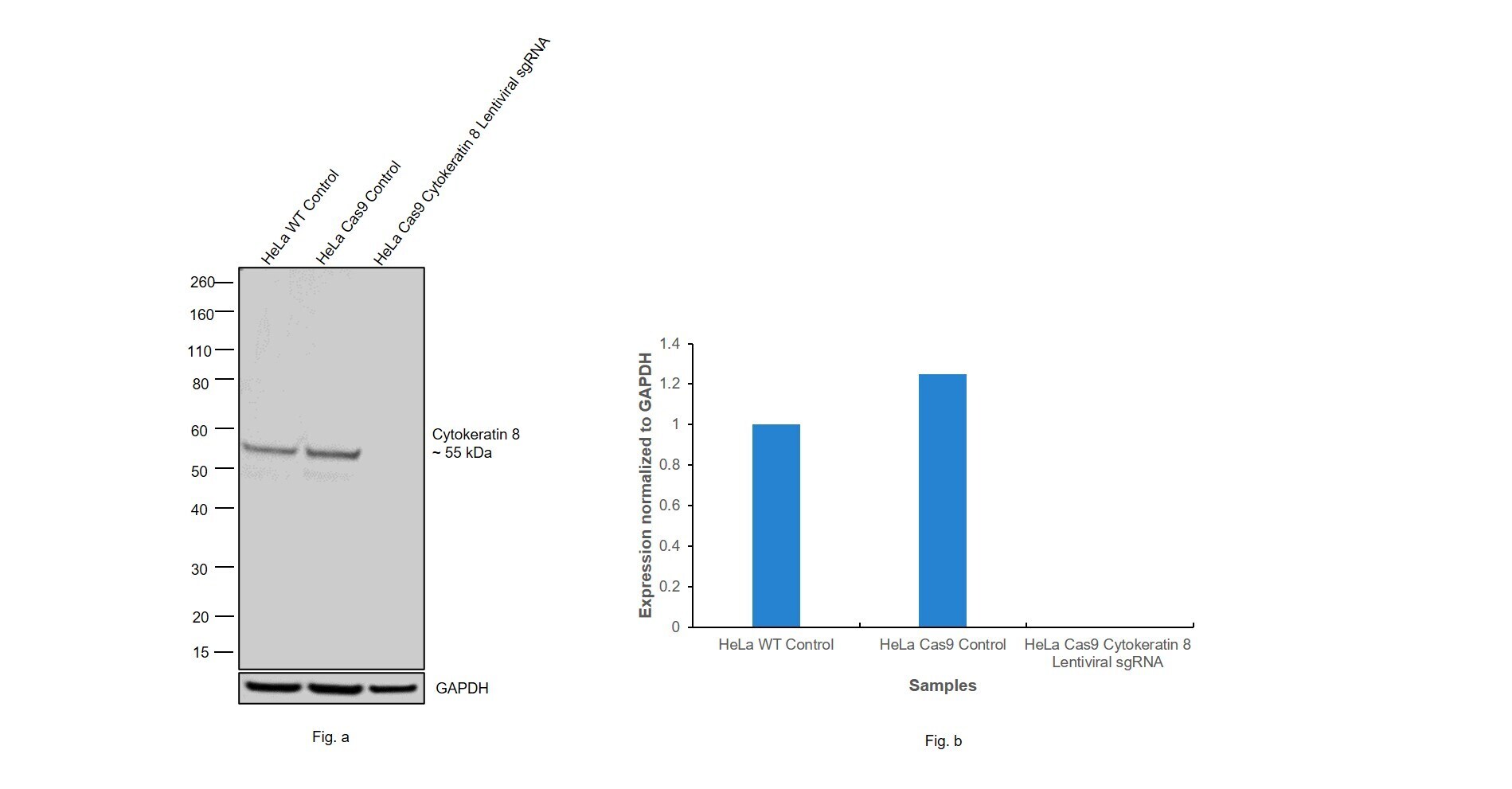
- CRISPR-Cas9 mediated genome editing ofCytokeratin 8 (as confirmed by next generation sequencing) was achieved by using LentiArray™ Lentiviral sgRNA (Product # A32042, AssayID CRISPR745510_LV) and LentiArray Cas9 Lentivirus (Product # A32064). Fig (a) Western blot analysis of Cytokeratin 8 was performed by loading 30 µg of HeLa Wild Type (Lane 1), HeLa Cas9 (Lane 2) and HeLa Cas9 cells transduced with Cytokeratin 8 Lentiviral sgRNA (Lane 3) whole cell extracts. The samples were electrophoresed using NuPAGE™ Novex™ 4-12% Bis-Tris Protein Gel (Product # NP0322BOX). Resolved proteins were then transferred onto a nitrocellulose membrane (Product # IB23001) by iBlot® 2 Dry Blotting System (Product # IB21001). The blot was probed with Anti-Cytokeratin 8 Polyclonal Antibody (Product # PA5-32469) using 1:1,000 dilution and Goat anti-Rabbit IgG (H+L) Superclonal™ Recombinant Secondary Antibody, HRP (Product # A27036 1:5,000 dilution).Chemiluminescent detection was performed using Novex® ECL Chemiluminescent Substrate Reagent Kit (Product # WP20005). A loss of signal in sgRNA transduced cells using the LentiArray™ CRISPR product line confirms that antibody is specific toCytokeratin 8 (Fig (b)).
- Submitted by
- Invitrogen Antibodies (provider)
- Main image

- Experimental details
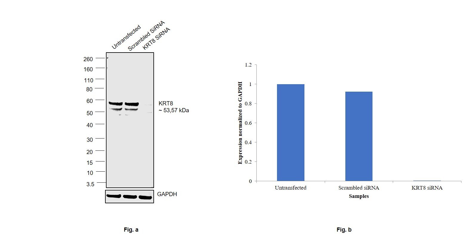
- Knockdown of Cytokeratin 8 was achieved by transfecting Caco-2 with Cytokeratin 8 specific siRNAs (Silencer® select Product # S7969, S7970). Western blot analysis (Fig. a) was performed using Whole cell extracts from the Cytokeratin 8 knockdown cells (lane 3), non-targeting scrambled siRNA transfected cells (lane 2) and untransfected cells (lane 1). The blot was probed with Cytokeratin 8 Polyclonal Antibody (Product # PA5-32469, 1:1000 dilution) and Goat anti-Rabbit IgG (H+L) Superclonal™ Recombinant Secondary Antibody, HRP (Product # A27036, 1:4000 dilution). Densitometric analysis of this western blot is shown in histogram (Fig. b). Decrease in signal upon siRNA mediated knock down confirms that antibody is specific to Cytokeratin 8.
- Submitted by
- Invitrogen Antibodies (provider)
- Main image
- Experimental details
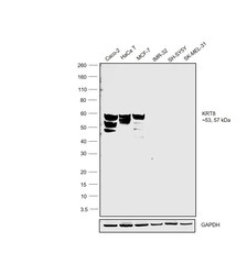
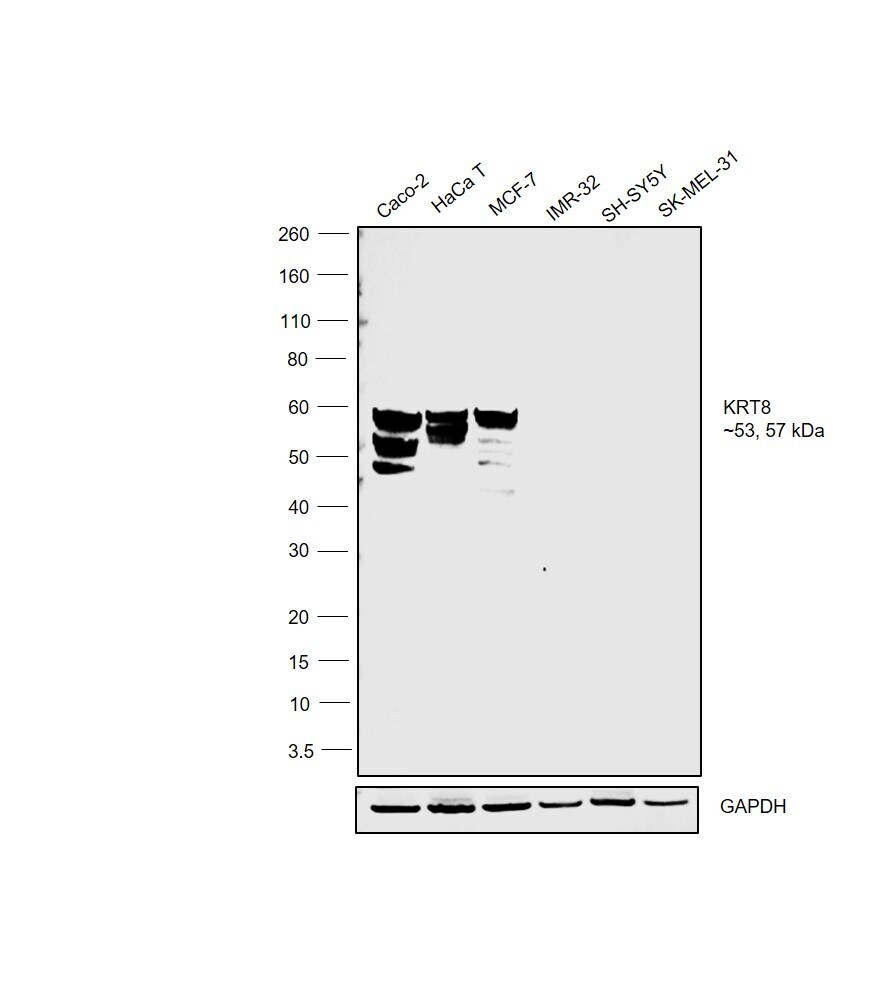
- Western blot was performed using Anti-Cytokeratin 8 Polyclonal Antibody(Product # PA5-32469) and a 53,57kDa band corresponding to Cytokeratin 8 was observed across the cell lines tested except IMR-32, SH-SY5Y and SK-MEL-32. Whole cell extracts (30 µg lysate) of Caco-2 (Lane 1), HaCaT (Lane 2), MCF7 (Lane 3), IMR-32 (Lane 4), SH-SY5Y (Lane 5) and SK-MEL-31 (Lane 6) electrophoresed using NuPAGE™ 4-12% Bis-Tris Protein Gel (Product # NP0321BOX). Resolved proteins were then transferred onto a Nitrocellulose membrane (Product # LC2002) by iBlot® 2 Dry Blotting System (Product # IB21001). The blot was probed with the primary antibody (1:1000 dilution) and detected by chemiluminescence with Goat anti-Rabbit IgG (H+L) Superclonal™ Recombinant Secondary Antibody, HRP (Product # A27036,1:4000 dilution) using the iBright FL 1000 (Product # A32752). Chemiluminescent detection was performed using Novex® ECL Chemiluminescent Substrate Reagent Kit (Product # WP20005).
Supportive validation
- Submitted by
- Invitrogen Antibodies (provider)
- Main image

- Experimental details
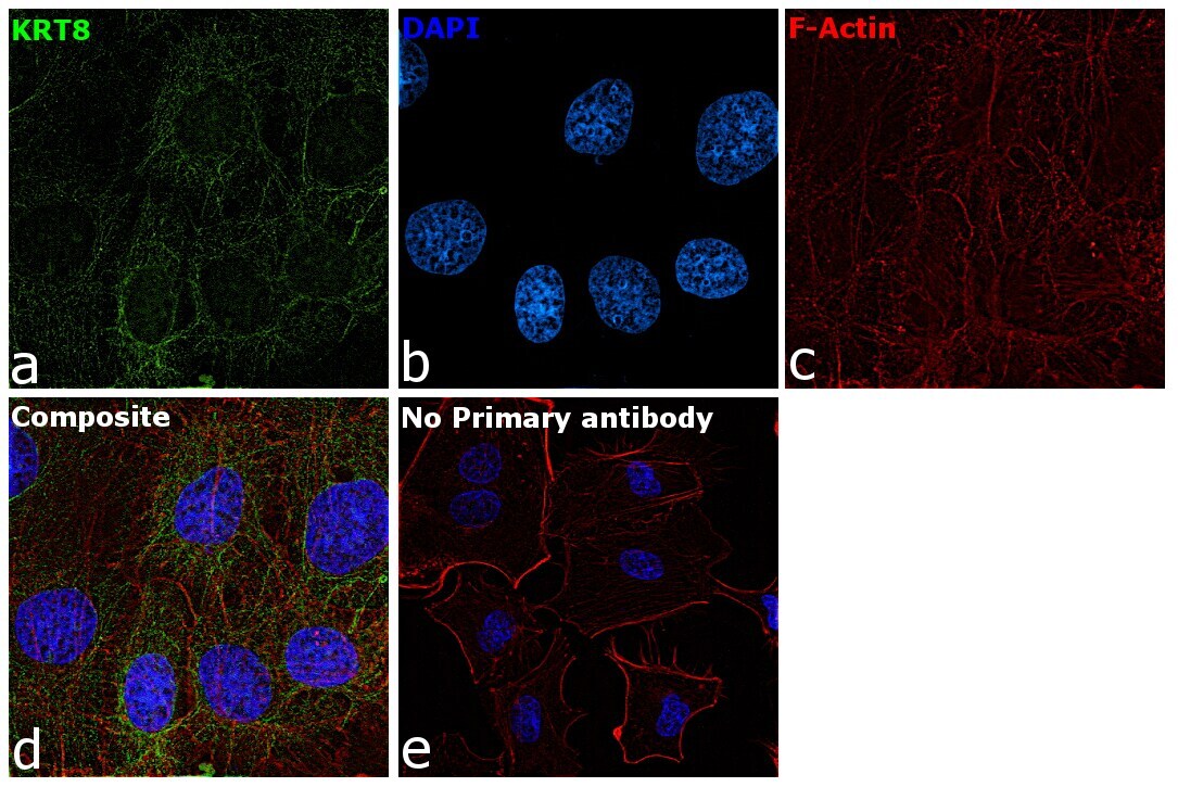
- Immunofluorescence analysis of Cytokeratin 8 was performed using 70% confluent log phase Caco-2 cells. The cells were fixed with ice-cold acetone at 4°C for 5 minutes, blocked with 2% BSA for 45 minutes at room temperature. The cells were labeled with Cytokeratin 8 Polyclonal Antibody (Product # PA5-32469) at 1:100 dilution in 0.1% BSA, incubated at 4 degree celsius overnight and then labeled with Goat anti-Rabbit IgG (H+L) Superclonal™ Recombinant Secondary Antibody, Alexa Fluor® 488 conjugate (Product # A27034), (1:2000 dilution), for 45 minutes at room temperature (Panel a: Green). Nuclei (Panel b:Blue) were stained with ProLong™ Diamond Antifade Mountant with DAPI (Product # P36962). F-actin (Panel c: Red) was stained with Rhodamine Phalloidin (Product # R415, 1:300 dilution). Panel d represents the merged image showing cytoskeletal localization. Panel e represents control cells with no primary antibody to assess background. The images were captured at 60X magnification.
Supportive validation
- Submitted by
- Invitrogen Antibodies (provider)
- Main image
- Experimental details
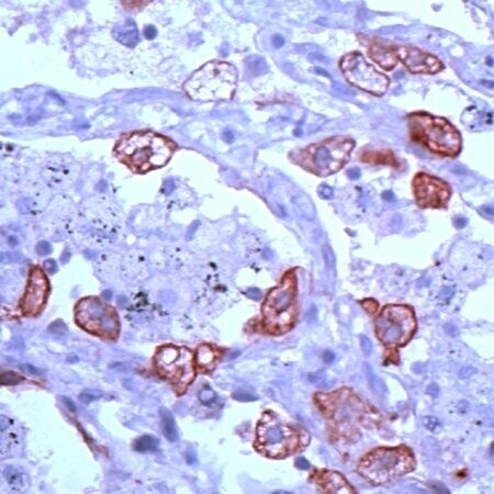
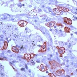
- Immunohistochemical analysis of Keratin 8 using anti-Keratin 8 Polyclonal Antibody (Product # PA5-32469) in Lung Cancer Tissue. The recommened dilution for this antibody in immunohistochemistry applications is 1:100.