 Explore
 Validate
 Learn
Learn Western blot
 Immunocytochemistry
Antibody data
- Antibody Data
- Antigen structure
- References [8]
- Comments [0]
- Validations
- Immunocytochemistry [2]
- Immunohistochemistry [3]
- Flow cytometry [1]
- Other assay [3]
Submit
Validation data
Reference
Comment
Report error
- Product number
- PA1-768 - Provider product page

- Provider
- Invitrogen Antibodies
- Product name
- VAMP4 Polyclonal Antibody
- Antibody type
- Polyclonal
- Antigen
- Recombinant full-length protein
- Description
- PA1-768 detects VAMP-4 from human and mouse samples. PA1-768 has been successfully used in Western blot and immunofluorescence procedures. By Western blot, this antibody detects an ~18 kDa protein representing VAMP-4 from PC3 cell extract. Immunofluorescent staining of VAMP-4 in CV-1 cells using PA1-768 results in a staining pattern consistent with VAMP-4 staining. Immunofluorescence staining with this antibody requires saponin permeabilization. The PA1-768 antigen is recombinant rat VAMP-4.
- Reactivity
- Human, Mouse, Rat
- Host
- Rabbit
- Isotype
- IgG
- Vial size
- 100 μg
- Concentration
- 1 mg/mL
- Storage
- -20°C, Avoid Freeze/Thaw Cycles
Submitted references SNAP23 deficiency causes severe brain dysplasia through the loss of radial glial cell polarity.
Control of insulin granule formation and function by the ABC transporters ABCG1 and ABCA1 and by oxysterol binding protein OSBP.
Vesicular transport system in myotubes: ultrastructural study and signposting with vesicle-associated membrane proteins.
Calcium-dependent activator protein for secretion 2 interacts with the class II ARF small GTPases and regulates dense-core vesicle trafficking.
An intracellular role for ABCG1-mediated cholesterol transport in the regulated secretory pathway of mouse pancreatic beta cells.
Interaction of calcium-dependent activator protein for secretion 1 (CAPS1) with the class II ADP-ribosylation factor small GTPases is required for dense-core vesicle trafficking in the trans-Golgi network.
VAMP2 marks quiescent satellite cells and myotubes, but not activated myoblasts.
The SNAREs vti1a and vti1b have distinct localization and SNARE complex partners.
Kunii M, Noguchi Y, Yoshimura SI, Kanda S, Iwano T, Avriyanti E, Atik N, Sato T, Sato K, Ogawa M, Harada A
The Journal of cell biology 2021 Jan 4;220(1)
The Journal of cell biology 2021 Jan 4;220(1)
Control of insulin granule formation and function by the ABC transporters ABCG1 and ABCA1 and by oxysterol binding protein OSBP.
Hussain SS, Harris MT, Kreutzberger AJB, Inouye CM, Doyle CA, Castle AM, Arvan P, Castle JD
Molecular biology of the cell 2018 May 15;29(10):1238-1257
Molecular biology of the cell 2018 May 15;29(10):1238-1257
Vesicular transport system in myotubes: ultrastructural study and signposting with vesicle-associated membrane proteins.
Tajika Y, Takahashi M, Khairani AF, Ueno H, Murakami T, Yorifuji H
Histochemistry and cell biology 2014 Apr;141(4):441-54
Histochemistry and cell biology 2014 Apr;141(4):441-54
Calcium-dependent activator protein for secretion 2 interacts with the class II ARF small GTPases and regulates dense-core vesicle trafficking.
Sadakata T, Sekine Y, Oka M, Itakura M, Takahashi M, Furuichi T
The FEBS journal 2012 Feb;279(3):384-94
The FEBS journal 2012 Feb;279(3):384-94
An intracellular role for ABCG1-mediated cholesterol transport in the regulated secretory pathway of mouse pancreatic beta cells.
Sturek JM, Castle JD, Trace AP, Page LC, Castle AM, Evans-Molina C, Parks JS, Mirmira RG, Hedrick CC
The Journal of clinical investigation 2010 Jul;120(7):2575-89
The Journal of clinical investigation 2010 Jul;120(7):2575-89
Interaction of calcium-dependent activator protein for secretion 1 (CAPS1) with the class II ADP-ribosylation factor small GTPases is required for dense-core vesicle trafficking in the trans-Golgi network.
Sadakata T, Shinoda Y, Sekine Y, Saruta C, Itakura M, Takahashi M, Furuichi T
The Journal of biological chemistry 2010 Dec 3;285(49):38710-9
The Journal of biological chemistry 2010 Dec 3;285(49):38710-9
VAMP2 marks quiescent satellite cells and myotubes, but not activated myoblasts.
Tajika Y, Takahashi M, Hino M, Murakami T, Yorifuji H
Acta histochemica et cytochemica 2010 Aug 27;43(4):107-14
Acta histochemica et cytochemica 2010 Aug 27;43(4):107-14
The SNAREs vti1a and vti1b have distinct localization and SNARE complex partners.
Kreykenbohm V, Wenzel D, Antonin W, Atlachkine V, von Mollard GF
European journal of cell biology 2002 May;81(5):273-80
European journal of cell biology 2002 May;81(5):273-80
No comments: Submit comment
Supportive validation
- Submitted by
- Invitrogen Antibodies (provider)
- Main image

- Experimental details
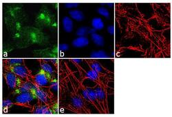
- Immunofluorescence analysis of VAMP4 was performed using 70% confluent log phase SH-SY5Y cells. The cells were fixed with 4% paraformaldehyde for 10 minutes, permeabilized with 0.1% Triton™ X-100 for 10 minutes, and blocked with 2% BSA for 1 hour at room temperature. The cells were labeled with VAMP4 Rabbit Polyclonal Antibody (Product # PA1-768) at 2 µg/mL in 0.1% BSA and incubated for 3 hours at room temperature and then labeled with Goat anti-Rabbit IgG (H+L) Superclonal™ Secondary Antibody, Alexa Fluor® 488 conjugate (Product # A27034) a dilution of 1:2000 for 45 minutes at room temperature (Panel a: green). Nuclei (Panel b: blue) were stained with SlowFade® Gold Antifade Mountant with DAPI (Product # S36938). F-actin (Panel c: red) was stained with Alexa Fluor® 555 Rhodamine Phalloidin (Product # R415, 1:300). Panel d represents the merged image showing cytoplasmic localization. Panel e shows the no primary antibody control. The images were captured at 60X magnification.

- Submitted by
- Invitrogen Antibodies (provider)
- Main image
- Experimental details
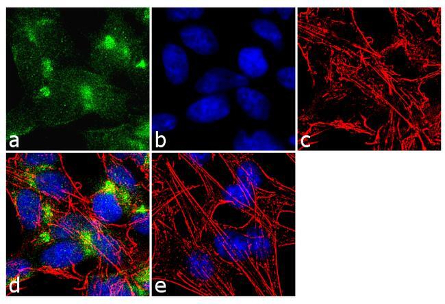
- Immunofluorescence analysis of VAMP4 was performed using 70% confluent log phase SH-SY5Y cells. The cells were fixed with 4% paraformaldehyde for 10 minutes, permeabilized with 0.1% Triton™ X-100 for 10 minutes, and blocked with 2% BSA for 1 hour at room temperature. The cells were labeled with VAMP4 Rabbit Polyclonal Antibody (Product # PA1-768) at 2 µg/mL in 0.1% BSA and incubated for 3 hours at room temperature and then labeled with Goat anti-Rabbit IgG (Heavy Chain) Superclonal™ Secondary Antibody, Alexa Fluor® 488 conjugate (Product # A27034) a dilution of 1:2000 for 45 minutes at room temperature (Panel a: green). Nuclei (Panel b: blue) were stained with SlowFade® Gold Antifade Mountant with DAPI (Product # S36938). F-actin (Panel c: red) was stained with Alexa Fluor® 555 Rhodamine Phalloidin (Product # R415, 1:300). Panel d represents the merged image showing cytoplasmic localization. Panel e shows the no primary antibody control. The images were captured at 60X magnification.
Supportive validation
- Submitted by
- Invitrogen Antibodies (provider)
- Main image
- Experimental details
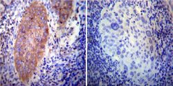
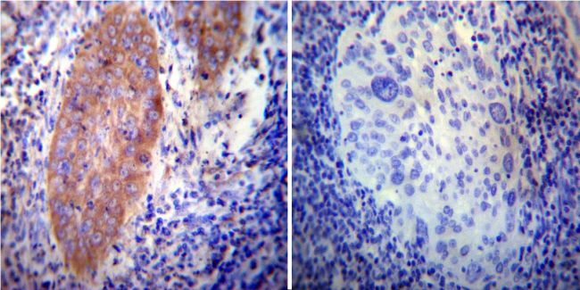
- Immunohistochemistry was performed on cancer biopsies of deparaffinized human Cervical carcinoma tissue. To expose target proteins, heat induced antigen retrieval was performed using 10mM sodium citrate (pH6.0) buffer, microwaved for 8-15 minutes. Following antigen retrieval tissues were blocked in 3% BSA-PBS for 30 minutes at room temperature. Tissues were then probed at a dilution of 1:200 with a rabbit polyclonal antibody recognizing VAMP4 (Product # PA1-768) or without primary antibody (negative control) overnight at 4°C in a humidified chamber. Tissues were washed extensively with PBST and endogenous peroxidase activity was quenched with a peroxidase suppressor. Detection was performed using a biotin-conjugated secondary antibody and SA-HRP, followed by colorimetric detection using DAB. Tissues were counterstained with hematoxylin and prepped for mounting.
- Submitted by
- Invitrogen Antibodies (provider)
- Main image
- Experimental details
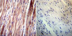
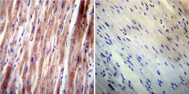
- Immunohistochemistry was performed on normal deparaffinized human Heart tissue. To expose target proteins, heat induced antigen retrieval was performed using 10mM sodium citrate (pH6.0) buffer, microwaved for 8-15 minutes. Following antigen retrieval tissues were blocked in 3% BSA-PBS for 30 minutes at room temperature. Tissues were then probed at a dilution of 1:200 with a rabbit polyclonal antibody recognizing VAMP4 (Product # PA1-768) or without primary antibody (negative control) overnight at 4°C in a humidified chamber. Tissues were washed extensively with PBST and endogenous peroxidase activity was quenched with a peroxidase suppressor. Detection was performed using a biotin-conjugated secondary antibody and SA-HRP, followed by colorimetric detection using DAB. Tissues were counterstained with hematoxylin and prepped for mounting.
- Submitted by
- Invitrogen Antibodies (provider)
- Main image
- Experimental details
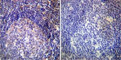
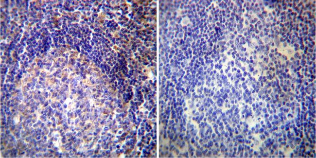
- Immunohistochemistry was performed on normal deparaffinized human Tonsil tissue. To expose target proteins, heat induced antigen retrieval was performed using 10mM sodium citrate (pH6.0) buffer, microwaved for 8-15 minutes. Following antigen retrieval tissues were blocked in 3% BSA-PBS for 30 minutes at room temperature. Tissues were then probed at a dilution of 1:200 with a rabbit polyclonal antibody recognizing VAMP4 (Product # PA1-768) or without primary antibody (negative control) overnight at 4°C in a humidified chamber. Tissues were washed extensively with PBST and endogenous peroxidase activity was quenched with a peroxidase suppressor. Detection was performed using a biotin-conjugated secondary antibody and SA-HRP, followed by colorimetric detection using DAB. Tissues were counterstained with hematoxylin and prepped for mounting.
Supportive validation
- Submitted by
- Invitrogen Antibodies (provider)
- Main image
- Experimental details
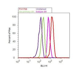
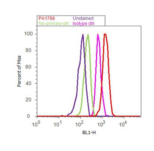
- Flow cytometry analysis of VAMP4 was done on SH-SY5Y cells. Cells were fixed with 70% ethanol for 10 minutes, permeabilized with 0.25% Triton™ X-100 for 20 minutes, and blocked with 5% BSA for 30 minutes at room temperature. Cells were labeled with VAMP4 Rabbit Polyclonal Antibody (PA1768, red histogram) or with rabbit isotype control (pink histogram) at 3-5 ug/million cells in 2.5% BSA. After incubation at room temperature for 2 hours, the cells were labeled with Alexa Fluor® 488 Goat Anti-Rabbit Secondary Antibody (A11008) at a dilution of 1:400 for 30 minutes at room temperature. The representative 10,000 cells were acquired and analyzed for each sample using an Attune® Acoustic Focusing Cytometer. The purple histogram represents unstained control cells and the green histogram represents no-primary-antibody control.
Supportive validation
- Submitted by
- Invitrogen Antibodies (provider)
- Main image

- Experimental details
- NULL
- Submitted by
- Invitrogen Antibodies (provider)
- Main image

- Experimental details
- NULL
- Submitted by
- Invitrogen Antibodies (provider)
- Main image

- Experimental details
- Figure S2. Knockdown of SNARE proteins in cultured RGCs. (A) Immunoblots showing each SNARE protein in the control (Ctrl) RGCs and RGCs with each SNARE protein depleted. (B and C) Cell aggregation assay of control RGCs and RGCs with each SNARE protein depleted and treated with or without 5 mM EGTA. The density of the aggregates was determined based on three independent experiments (C). Scale bar, 100 um. (D) N-cadherin (Ncad; green) and VAMP3 (upper, red), VAMP4 (middle, red), or VAMP5 (lower, red) stained with DAPI (blue) in the control and VAMP3-, VAMP4-, or VAMP5-depleted RGCs. Scale bar, 10 um. The bar graph represents the means +- SEM. Significance was calculated using two-tailed paired Student's t tests. Statistical significance is indicated by *P < 0.05 or **P < 0.01.
