Explore
 Validate
 Learn
Learn Western blot
 Immunohistochemistry
Antibody data
- Antibody Data
- Antigen structure
- References [38]
- Comments [0]
- Validations
- Immunohistochemistry [1]
Submit
Validation data
Reference
Comment
Report error
- Product number
- HPA000288 - Provider product page

- Provider
- Atlas Antibodies
- Proper citation
- Atlas Antibodies Cat#HPA000288, RRID:AB_1078160
- Product name
- Anti-ACE2
- Antibody type
- Polyclonal
- Description
- Polyclonal Antibody against Human ACE2, Gene description: angiotensin I converting enzyme 2, Validated applications: IHC, WB, Uniprot ID: Q9BYF1, Storage: Store at +4°C for short term storage. Long time storage is recommended at -20°C.
- Reactivity
- Human
- Host
- Rabbit
- Conjugate
- Unconjugated
- Isotype
- IgG
- Vial size
- 100 µl
- Concentration
- 0.1 mg/ml
- Storage
- Store at +4°C for short term storage. Long time storage is recommended at -20°C.
- Handling
- The antibody solution should be gently mixed before use.
Submitted references Neuronal progenitors of the dentate gyrus express the SARS-CoV-2 cell receptor during migration in the developing human hippocampus
Myocardial Angiotensin-Converting Enzyme 2 Protein Expression in Ischemic Heart Failure
Glycosylated extracellular mucin domains protect against SARS-CoV-2 infection at the respiratory surface
SARS-CoV-2 replicates and displays oncolytic properties in clear cell and papillary renal cell carcinoma.
Regulation of angiotensin-converting enzyme 2 isoforms by type 2 inflammation and viral infection in human airway epithelium
Higher angiotensin-converting enzyme 2 (ACE2) levels in the brain of individuals with Alzheimer’s disease
Spike protein of SARS-CoV-2 suppresses gonadotrophin secretion from bovine anterior pituitaries
Fulminant Myocarditis 24 Days after Coronavirus Disease Messenger Ribonucleic Acid Vaccination
Angiotensin-converting enzyme 2 in peripheral lung club cells modulates the susceptibility to SARS-CoV-2 in chronic obstructive pulmonary disease
ACE2 Protein Expression During Childhood, Adolescence, and Early Adulthood
Ultrastructural insight into SARS-CoV-2 entry and budding in human airway epithelium
Comprehensive Analysis of Disease Pathology in Immunocompetent and Immunocompromised Hosts following Pulmonary SARS-CoV-2 Infection
Live imaging of SARS-CoV-2 infection in mice reveals that neutralizing antibodies require Fc function for optimal efficacy
Acute Respiratory Distress in Aged, SARS-CoV-2–Infected African Green Monkeys but Not Rhesus Macaques
Secukinumab lowers expression of ACE2 in affected skin of patients with psoriasis
Epithelial response to IFN‐γ promotes SARS‐CoV‐2 infection
Unexpected tumor reduction in metastatic colorectal cancer patients during SARS-Cov-2 infection
Ovulatory upregulation of angiotensin-converting enzyme 2, a receptor for SARS-CoV-2, in dominant follicles of the human ovary
Elevation in viral entry genes and innate immunity compromise underlying increased infectivity and severity of COVID-19 in cancer patients
Effects of Angiotensin II Type 1A Receptor on ACE2, Neprilysin and KIM-1 in Two Kidney One Clip (2K1C) Model of Renovascular Hypertension
Nasal delivery of single-domain antibody improves symptoms of SARS-CoV-2 infection in an animal model.
An organoid-derived bronchioalveolar model for SARS-CoV-2 infection of human alveolar type II-like cells.
ACE2: Evidence of role as entry receptor for SARS-CoV-2 and implications in comorbidities
A profiling analysis on the receptor ACE2 expression reveals the potential risk of different type of cancers vulnerable to SARS-CoV-2 infection
Effect of hyperglycemia and rosiglitazone on renal and urinary neprilysin indb/dbdiabetic mice
A bioinformatics analysis on the potential role of ACE2 in cardiac impairment of patients with coronavirus disease 2019
Expressions and significances of the angiotensin-converting enzyme 2 gene, the receptor of SARS-CoV-2 for COVID-19
Expression of SARS-CoV-2 Entry Factors in the Pancreas of Normal Organ Donors and Individuals with COVID-19
Bioinformatic Analysis of Correlation between Immune Infiltration and COVID-19 in Cancer Patients
Bioinformatic characterization of angiotensin-converting enzyme 2, the entry receptor for SARS-CoV-2
ACE2 localizes to the respiratory cilia and is not increased by ACE inhibitors or ARBs
The protein expression profile of ACE2 in human tissues.
SARS-CoV-2 Cell Entry Factors ACE2 and TMPRSS2 Are Expressed in the Microvasculature and Ducts of Human Pancreas but Are Not Enriched in β Cells.
Variance decomposition of protein profiles from antibody arrays using a longitudinal twin model
Gene Expression Profiling of Metaplastic Lineages Identifies CDH17 as a Prognostic Marker in Early Stage Gastric Cancer
Hernandez-Lopez J, Hernandez-Medina C, Medina-Corvalan C, Rodenas M, Francisca A, Perez-Garcia C, Echevarria D, Carratala F, Geijo-Barrientos E, Martinez S
Cellular and Molecular Life Sciences 2023;80(6)
Cellular and Molecular Life Sciences 2023;80(6)
Myocardial Angiotensin-Converting Enzyme 2 Protein Expression in Ischemic Heart Failure
Siratavičiūtė V, Pangonytė D, Utkienė L, Jusienė L, Marcinkevičienė J, Stanionienė Z, Radikė R
International Journal of Molecular Sciences 2023;24(24):17145
International Journal of Molecular Sciences 2023;24(24):17145
Glycosylated extracellular mucin domains protect against SARS-CoV-2 infection at the respiratory surface
Frieman M, Chatterjee M, Huang L, Mykytyn A, Wang C, Lamers M, Westendorp B, Wubbolts R, van Putten J, Bosch B, Haagmans B, Strijbis K
PLOS Pathogens 2023;19(8):e1011571
PLOS Pathogens 2023;19(8):e1011571
SARS-CoV-2 replicates and displays oncolytic properties in clear cell and papillary renal cell carcinoma.
Choong OK, Jakobsson R, Bergdahl AG, Brunet S, Kärmander A, Waldenström J, Arvidsson Y, Altiparmak G, Nilsson JA, Karlsson J, Nyström K, Johansson ME
PloS one 2023;18(1):e0279578
PloS one 2023;18(1):e0279578
Regulation of angiotensin-converting enzyme 2 isoforms by type 2 inflammation and viral infection in human airway epithelium
Stocker N, Radzikowska U, Wawrzyniak P, Tan G, Huang M, Ding M, Akdis C, Sokolowska M
Mucosal Immunology 2023;16(1):5-16
Mucosal Immunology 2023;16(1):5-16
Higher angiotensin-converting enzyme 2 (ACE2) levels in the brain of individuals with Alzheimer’s disease
Reveret L, Leclerc M, Emond V, Tremblay C, Loiselle A, Bourassa P, Bennett D, Hébert S, Calon F
Acta Neuropathologica Communications 2023;11(1)
Acta Neuropathologica Communications 2023;11(1)
Louise R, Manon L, Vincent E, Andréanne L, Philippe B, Cyntia T, Bennett D, Sébastien H, Frédéric C
2023
2023
Spike protein of SARS-CoV-2 suppresses gonadotrophin secretion from bovine anterior pituitaries
ABDILLAH D, KEREILWE O, FERDOUSY R, SAITO R, KADOKAWA H
Journal of Reproduction and Development 2022;68(2):152-159
Journal of Reproduction and Development 2022;68(2):152-159
Fulminant Myocarditis 24 Days after Coronavirus Disease Messenger Ribonucleic Acid Vaccination
Kawano H, Motokawa T, Kurohama H, Okano S, Akashi R, Yonekura T, Ikeda S, Izumikawa K, Maemura K
Internal Medicine 2022;61(15):2319-2325
Internal Medicine 2022;61(15):2319-2325
Angiotensin-converting enzyme 2 in peripheral lung club cells modulates the susceptibility to SARS-CoV-2 in chronic obstructive pulmonary disease
Peng Y, Wang Z, Chen S, Xu A, Fang Z, Sun J, Zhou Z, Hou X, Cen L, Ma J, Zhao J, Guan W, Wang D, Zhong N
American Journal of Physiology-Lung Cellular and Molecular Physiology 2022;322(5):L712-L721
American Journal of Physiology-Lung Cellular and Molecular Physiology 2022;322(5):L712-L721
ACE2 Protein Expression During Childhood, Adolescence, and Early Adulthood
Schurink B, Roos E, Vos W, Breur M, van der Valk P, Bugiani M
Pediatric and Developmental Pathology 2022;25(4):404-408
Pediatric and Developmental Pathology 2022;25(4):404-408
Ultrastructural insight into SARS-CoV-2 entry and budding in human airway epithelium
Pinto A, Rai R, Brown J, Griffin P, Edgar J, Shah A, Singanayagam A, Hogg C, Barclay W, Futter C, Burgoyne T
Nature Communications 2022;13(1)
Nature Communications 2022;13(1)
Comprehensive Analysis of Disease Pathology in Immunocompetent and Immunocompromised Hosts following Pulmonary SARS-CoV-2 Infection
Ramasamy S, Kolloli A, Kumar R, Husain S, Soteropoulos P, Chang T, Subbian S
Biomedicines 2022;10(6):1343
Biomedicines 2022;10(6):1343
Live imaging of SARS-CoV-2 infection in mice reveals that neutralizing antibodies require Fc function for optimal efficacy
Ullah I, Prévost J, Ladinsky M, Stone H, Lu M, Anand S, Beaudoin-Bussières G, Symmes K, Benlarbi M, Ding S, Gasser R, Fink C, Chen Y, Tauzin A, Goyette G, Bourassa C, Medjahed H, Mack M, Chung K, Wilen C, Dekaban G, Dikeakos J, Bruce E, Kaufmann D, Stamatatos L, McGuire A, Richard J, Pazgier M, Bjorkman P, Mothes W, Finzi A, Kumar P, Uchil P
Immunity 2021;54(9):2143-2158.e15
Immunity 2021;54(9):2143-2158.e15
Acute Respiratory Distress in Aged, SARS-CoV-2–Infected African Green Monkeys but Not Rhesus Macaques
Blair R, Vaccari M, Doyle-Meyers L, Roy C, Russell-Lodrigue K, Fahlberg M, Monjure C, Beddingfield B, Plante K, Plante J, Weaver S, Qin X, Midkiff C, Lehmicke G, Golden N, Threeton B, Penney T, Allers C, Barnes M, Pattison M, Datta P, Maness N, Birnbaum A, Fischer T, Bohm R, Rappaport J
The American Journal of Pathology 2021;191(2):274-282
The American Journal of Pathology 2021;191(2):274-282
Secukinumab lowers expression of ACE2 in affected skin of patients with psoriasis
Krueger J, Murrell D, Garcet S, Navrazhina K, Lee P, Muscianisi E, Blauvelt A
Journal of Allergy and Clinical Immunology 2021;147(3):1107-1109.e2
Journal of Allergy and Clinical Immunology 2021;147(3):1107-1109.e2
Epithelial response to IFN‐γ promotes SARS‐CoV‐2 infection
Heuberger J, Trimpert J, Vladimirova D, Goosmann C, Lin M, Schmuck R, Mollenkopf H, Brinkmann V, Tacke F, Osterrieder N, Sigal M
EMBO Molecular Medicine 2021;13(4)
EMBO Molecular Medicine 2021;13(4)
Unexpected tumor reduction in metastatic colorectal cancer patients during SARS-Cov-2 infection
Ottaiano A, Scala S, D’Alterio C, Trotta A, Bello A, Rea G, Picone C, Santorsola M, Petrillo A, Nasti G
Therapeutic Advances in Medical Oncology 2021;13
Therapeutic Advances in Medical Oncology 2021;13
Ovulatory upregulation of angiotensin-converting enzyme 2, a receptor for SARS-CoV-2, in dominant follicles of the human ovary
Choi Y, Jeon H, Brännström M, Akin J, Curry T, Jo M
Fertility and Sterility 2021;116(6):1631-1640
Fertility and Sterility 2021;116(6):1631-1640
Elevation in viral entry genes and innate immunity compromise underlying increased infectivity and severity of COVID-19 in cancer patients
Kwan J, Lin L, Bell R, Bruce J, Richardson C, Pugh T, Liu F
Scientific Reports 2021;11(1)
Scientific Reports 2021;11(1)
Effects of Angiotensin II Type 1A Receptor on ACE2, Neprilysin and KIM-1 in Two Kidney One Clip (2K1C) Model of Renovascular Hypertension
Alawi L, Dhakal S, Emberesh S, Sawant H, Hosawi A, Thanekar U, Grobe N, Elased K
Frontiers in Pharmacology 2021;11
Frontiers in Pharmacology 2021;11
Nasal delivery of single-domain antibody improves symptoms of SARS-CoV-2 infection in an animal model.
Haga K, Takai-Todaka R, Matsumura Y, Song C, Takano T, Tojo T, Nagami A, Ishida Y, Masaki H, Tsuchiya M, Ebisudani T, Sugimoto S, Sato T, Yasuda H, Fukunaga K, Sawada A, Nemoto N, Murata K, Morimoto T, Katayama K
PLoS pathogens 2021 Oct;17(10):e1009542
PLoS pathogens 2021 Oct;17(10):e1009542
An organoid-derived bronchioalveolar model for SARS-CoV-2 infection of human alveolar type II-like cells.
Lamers MM, van der Vaart J, Knoops K, Riesebosch S, Breugem TI, Mykytyn AZ, Beumer J, Schipper D, Bezstarosti K, Koopman CD, Groen N, Ravelli RBG, Duimel HQ, Demmers JAA, Verjans GMGM, Koopmans MPG, Muraro MJ, Peters PJ, Clevers H, Haagmans BL
The EMBO journal 2021 Mar 1;40(5):e105912
The EMBO journal 2021 Mar 1;40(5):e105912
ACE2: Evidence of role as entry receptor for SARS-CoV-2 and implications in comorbidities
Zamorano Cuervo N, Grandvaux N
eLife 2020;9
eLife 2020;9
A profiling analysis on the receptor ACE2 expression reveals the potential risk of different type of cancers vulnerable to SARS-CoV-2 infection
Dai Y, Hu F, Li H, Huang H, Wang D, Liang Y
Annals of Translational Medicine 2020;8(7):481-481
Annals of Translational Medicine 2020;8(7):481-481
Effect of hyperglycemia and rosiglitazone on renal and urinary neprilysin indb/dbdiabetic mice
Alawi L, Emberesh S, Owuor B, Chodavarapu H, Fadnavis R, El‐Amouri S, Elased K
Physiological Reports 2020;8(3)
Physiological Reports 2020;8(3)
A bioinformatics analysis on the potential role of ACE2 in cardiac impairment of patients with coronavirus disease 2019
Wang W, Lv S, Gan W, Zeng Z, Yang M
Annals of Translational Medicine 2020;8(21):1403-1403
Annals of Translational Medicine 2020;8(21):1403-1403
Expressions and significances of the angiotensin-converting enzyme 2 gene, the receptor of SARS-CoV-2 for COVID-19
Fu J, Zhou B, Zhang L, Balaji K, Wei C, Liu X, Chen H, Peng J, Fu J
Molecular Biology Reports 2020;47(6):4383-4392
Molecular Biology Reports 2020;47(6):4383-4392
Expression of SARS-CoV-2 Entry Factors in the Pancreas of Normal Organ Donors and Individuals with COVID-19
Kusmartseva I, Wu W, Syed F, Van Der Heide V, Jorgensen M, Joseph P, Tang X, Candelario-Jalil E, Yang C, Nick H, Harbert J, Posgai A, Paulsen J, Lloyd R, Cechin S, Pugliese A, Campbell-Thompson M, Vander Heide R, Evans-Molina C, Homann D, Atkinson M
Cell Metabolism 2020;32(6):1041-1051.e6
Cell Metabolism 2020;32(6):1041-1051.e6
Bioinformatic Analysis of Correlation between Immune Infiltration and COVID-19 in Cancer Patients
Huang X, He C, Hua X, Kan A, Sun S, Wang J, Li S
International Journal of Biological Sciences 2020;16(13):2464-2476
International Journal of Biological Sciences 2020;16(13):2464-2476
Bioinformatic characterization of angiotensin-converting enzyme 2, the entry receptor for SARS-CoV-2
Bader M, Barker H, Parkkila S
PLOS ONE 2020;15(10):e0240647
PLOS ONE 2020;15(10):e0240647
ACE2 localizes to the respiratory cilia and is not increased by ACE inhibitors or ARBs
Lee I, Nakayama T, Wu C, Goltsev Y, Jiang S, Gall P, Liao C, Shih L, Schürch C, McIlwain D, Chu P, Borchard N, Zarabanda D, Dholakia S, Yang A, Kim D, Chen H, Kanie T, Lin C, Tsai M, Phillips K, Kim R, Overdevest J, Tyler M, Yan C, Lin C, Lin Y, Bau D, Tsay G, Patel Z, Tsou Y, Tzankov A, Matter M, Tai C, Yeh T, Hwang P, Nolan G, Nayak J, Jackson P
Nature Communications 2020;11(1)
Nature Communications 2020;11(1)
The protein expression profile of ACE2 in human tissues.
Hikmet F, Méar L, Edvinsson Å, Micke P, Uhlén M, Lindskog C
Molecular systems biology 2020 Jul;16(7):e9610
Molecular systems biology 2020 Jul;16(7):e9610
SARS-CoV-2 Cell Entry Factors ACE2 and TMPRSS2 Are Expressed in the Microvasculature and Ducts of Human Pancreas but Are Not Enriched in β Cells.
Coate KC, Cha J, Shrestha S, Wang W, Gonçalves LM, Almaça J, Kapp ME, Fasolino M, Morgan A, Dai C, Saunders DC, Bottino R, Aramandla R, Jenkins R, Stein R, Kaestner KH, Vahedi G, HPAP Consortium, Brissova M, Powers AC
Cell metabolism 2020 Dec 1;32(6):1028-1040.e4
Cell metabolism 2020 Dec 1;32(6):1028-1040.e4
Lee I, Nakayama T, Wu C, Goltsev Y, Jiang S, Gall P, Liao C, Shih L, Schürch C, McIlwain D, Chu P, Borchard N, Zarabanda D, Dholakia S, Yang A, Kim D, Kanie T, Lin C, Tsai M, Phillips K, Kim R, Overdevest J, Tyler M, Yan C, Lin C, Lin Y, Bau D, Tsay G, Patel Z, Tsou Y, Tai C, Yeh T, Hwang P, Nolan G, Nayak J, Jackson P
2020
2020
Duijf P
2020
2020
Variance decomposition of protein profiles from antibody arrays using a longitudinal twin model
Kato B, Nicholson G, Neiman M, Rantalainen M, Holmes C, Barrett A, Uhlén M, Nilsson P, Spector T, Schwenk J
Proteome Science 2011;9(1):73
Proteome Science 2011;9(1):73
Gene Expression Profiling of Metaplastic Lineages Identifies CDH17 as a Prognostic Marker in Early Stage Gastric Cancer
Lee H, Nam K, Park H, Kim M, LaFleur B, Aburatani H, Yang H, Kim W, Goldenring J
Gastroenterology 2010;139(1):213-225.e3
Gastroenterology 2010;139(1):213-225.e3
No comments: Submit comment
Supportive validation
- Submitted by
- Atlas Antibodies (provider)
- Enhanced method
- Orthogonal validation
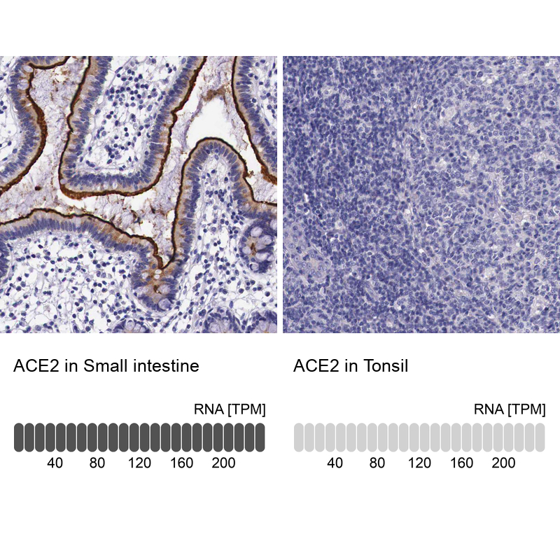
- Main image
- Experimental details
- Immunohistochemistry analysis in human small intestine and tonsil tissues using HPA000288 antibody. Corresponding ACE2 RNA-seq data are presented for the same tissues.
- Sample type
- Human
- Protocol
- Protocol