 Explore
 Validate
 Learn
Learn Western blot
Antibody data
- Antibody Data
- Antigen structure
- References [1]
- Comments [0]
- Validations
- Western blot [5]
- Immunocytochemistry [2]
- Immunohistochemistry [7]
Submit
Validation data
Reference
Comment
Report error
- Product number
- PA5-20040 - Provider product page

- Provider
- Invitrogen Antibodies
- Product name
- ACE2 Polyclonal Antibody
- Antibody type
- Polyclonal
- Antigen
- Synthetic peptide
- Description
- In Western blot applications, this antibody detects a band at ~90kDa. A suggested positive control is human kidney tissue lysate.
- Concentration
- 1 mg/mL
Submitted references Epstein-Barr Virus Lytic Replication Induces ACE2 Expression and Enhances SARS-CoV-2 Pseudotyped Virus Entry in Epithelial Cells.
Verma D, Church TM, Swaminathan S
Journal of virology 2021 Jun 10;95(13):e0019221
Journal of virology 2021 Jun 10;95(13):e0019221
No comments: Submit comment
Supportive validation
- Submitted by
- Invitrogen Antibodies (provider)
- Main image
- Experimental details
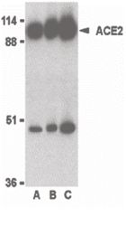
- Western blot analysis of human kidney lysate using a ACE2 polyclonal antibody (Product # PA5-20040) at 0.5 (lane A), 1 (lane B), and 2 (lane C) µg/mL.
- Submitted by
- Invitrogen Antibodies (provider)
- Main image

- Experimental details
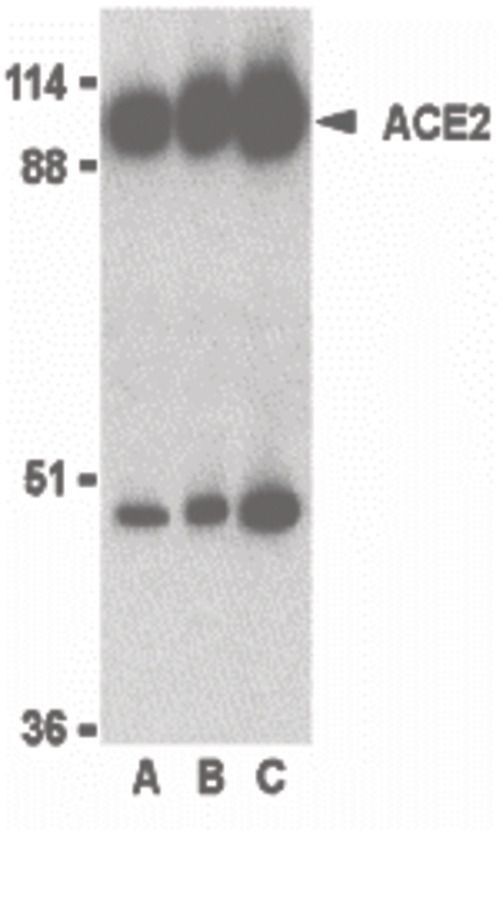
- Western Blot Validation in Human Tissues and Cell Line. Loading: 15 µg of lysates per lane. Antibodies: ACE2 Polyclonal Antibody (Product # PA5-20040) (2 µg/mL), 1h incubation at RT in 0.05 NFDM/TBST. Secondary: Goat anti-rabbit IgG HRP conjugate at 1:10,000 dilution.

- Submitted by
- Invitrogen Antibodies (provider)
- Main image
- Experimental details
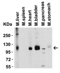
- Western Blot Validation in Mouse Tissues. Loading: 15 µg of lysates per lane. Antibodies: ACE2 Polyclonal Antibody (Product # PA5-20040) (2 µg/mL), 1h incubation at RT in 0.05 NFDM/TBST. Secondary: Goat anti-rabbit IgG HRP conjugate at 1:10,000 dilution.

- Submitted by
- Invitrogen Antibodies (provider)
- Main image
- Experimental details
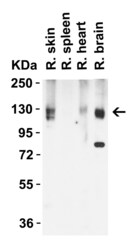
- Western Blot Validation in Rat Tissues. Loading: 15 µg of lysates per lane. Antibodies: ACE2 Polyclonal Antibody (Product # PA5-20040) (2 µg/mL), 1h incubation at RT in 0.05 NFDM/TBST. Secondary: Goat anti-rabbit IgG HRP conjugate at 1:10,000 dilution.

- Submitted by
- Invitrogen Antibodies (provider)
- Main image

- Experimental details
- Western Blot was performed using Anti-ACE2 Polyclonal Antibody (Product # PA5-20040) and a 90 kDa band corresponding to Angiotensin-converting enzyme 2 was observed across cell lines tested. Whole cell extracts (30 µg lysate) of MDA-MB-231(Lane 1), MCF7 (Lane 2),HCT 116 (Lane 3), Caco-2 (Lane 4), A549 (Lane 5), A549 treated with TGFb1 (10 ng/mL, 72 Hrs) (Lane 6) were electrophoresed using NuPAGE™ 4-12% Bis-Tris Protein Gel (Product # NP0322BOX). Resolved proteins were then transferred onto a nitrocellulose membrane (Product # IB23001) by iBlot® 2 Dry Blotting System (Product # IB21001). The blot was probed with the primary antibody (1:1000 Dilution) and detected by chemiluminescence with Goat anti-Rabbit IgG (H+L) Superclonal™ Recombinant Secondary Antibody, HRP (Product # A27036, 1:4000 dilution) using the iBright FL 1000 (Product # A32752). Chemiluminescent detection was performed using Novex® ECL Chemiluminescent Substrate Reagent Kit (Product # WP20005).Expression of SIAH1 was found to be higher in MCF-7,Caco-2 and A549 treated with TGFb1 (10 ng/mL, 72 Hrs) as compared to MDA-MB-231 and HCT 116.

Supportive validation
- Submitted by
- Invitrogen Antibodies (provider)
- Main image
- Experimental details
- Immunofluorescent analysis of human kidney cells using a ACE2 polyclonal antibody (Product # PA5-20040) at a 10 µg/mL dilution.

- Submitted by
- Invitrogen Antibodies (provider)
- Main image
- Experimental details
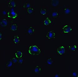
- Immunofluorescent analysis of 4% paraformaldehyde-fixed Caco2 cells labeling ACE2 with ACE2 Polyclonal Antibody (Product # PA5-20040) at 5 µg/mL, followed by goat anti-rabbit IgG secondary antibody at 1:500 dilution (green) and DAPI staining (blue). Image showing membrane staining on Caco2 cells.

Supportive validation
- Submitted by
- Invitrogen Antibodies (provider)
- Main image

- Experimental details
- Immunofluorescent analysis of 4% paraformaldehyde-fixed rat lung tissue labeling ACE-2 with ACE2 Polyclonal Antibody (Product # PA5-20040) at 20 µg/mL, followed by goat anti-rabbit IgG secondary antibody at 1:500 dilution (green) and DAPI staining (blue).

- Submitted by
- Invitrogen Antibodies (provider)
- Main image

- Experimental details
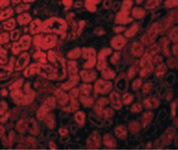
- Immunofluorescent analysis of 4% paraformaldehyde-fixed human kidney cells labeling ACE2 with ACE2 Polyclonal Antibody (Product # PA5-20040) at 10 µg/mL, followed by goat anti-rabbit IgG secondary antibody at 1:500 dilution (red).

- Submitted by
- Invitrogen Antibodies (provider)
- Main image

- Experimental details
- Immunofluorescent analysis of 4% paraformaldehyde-fixed human lung tissue labeling ACE-2 with ACE2 Polyclonal Antibody (Product # PA5-20040) at 20 µg/mL, followed by goat anti-rabbit IgG secondary antibody at 1:500 dilution (green) and DAPI staining (blue).

- Submitted by
- Invitrogen Antibodies (provider)
- Main image

- Experimental details
- Immunofluorescent analysis of 4% paraformaldehyde-fixed human testis tissue labeling ACE-2 with ACE2 Polyclonal Antibody (Product # PA5-20040) at 20 µg/mL, followed by goat anti-rabbit IgG secondary antibody at 1:500 dilution (green) and DAPI staining (blue).

- Submitted by
- Invitrogen Antibodies (provider)
- Main image

- Experimental details
- Immunofluorescent analysis of 4% paraformaldehyde-fixed mouse lung tissue labeling ACE-2 with ACE2 Polyclonal Antibody (Product # PA5-20040) at 20 µg/mL, followed by goat anti-rabbit IgG secondary antibody at 1:500 dilution (green) and DAPI staining (blue).

- Submitted by
- Invitrogen Antibodies (provider)
- Main image

- Experimental details
- Immunofluorescent analysis of 4% paraformaldehyde-fixed rat lung tissue labeling ACE-2 with ACE2 Polyclonal Antibody (Product # PA5-20040) at 20 µg/mL, followed by goat anti-rabbit IgG secondary antibody at 1:500 dilution (green) and DAPI staining (blue).

- Submitted by
- Invitrogen Antibodies (provider)
- Main image

- Experimental details
- Immunohistochemical analysis of paraffin-embedded human kidney tissue using ACE2 Polyclonal Antibody (Product # PA5-20040) at 2 µg/mL. Tissue was fixed with formaldehyde and blocked with 0.1 serum for 1 h at RT; antigen retrieval was by heat mediation with a citrate buffer (pH6). Samples were incubated with primary antibody overnight at 4˚C. A goat anti-rabbit IgG H&L (HRP) at 1/250 was used as secondary. Counter stained with Hematoxylin.
