Explore
 Validate
 Learn
Learn Western blot
 Immunocytochemistry
Antibody data
- Antibody Data
- Antigen structure
- References [2]
- Comments [0]
- Validations
- Immunocytochemistry [7]
- Immunohistochemistry [5]
- Other assay [2]
Submit
Validation data
Reference
Comment
Report error
- Product number
- PA5-20046 - Provider product page

- Provider
- Invitrogen Antibodies
- Product name
- ACE2 Polyclonal Antibody
- Antibody type
- Polyclonal
- Antigen
- Synthetic peptide
- Description
- In Western blot applications, this antibody detects a band at ~90kDa. A suggested positive control is human kidney tissue lysate. PA5-20046 can be used with blocking peptide PEP-0165.
- Reactivity
- Human
- Host
- Rabbit
- Isotype
- IgG
- Vial size
- 100 μg
- Concentration
- 1 mg/mL
- Storage
- Maintain refrigerated at 2-8°C for up to 3 months. For long term storage store at -20°C
Submitted references Differential Effect of SARS-CoV-2 Spike Glycoprotein 1 on Human Bronchial and Alveolar Lung Mucosa Models: Implications for Pathogenicity.
Paradoxical effects of cigarette smoke and COPD on SARS-CoV-2 infection and disease.
Rahman M, Irmler M, Keshavan S, Introna M, Beckers J, Palmberg L, Johanson G, Ganguly K, Upadhyay S
Viruses 2021 Dec 17;13(12)
Viruses 2021 Dec 17;13(12)
Paradoxical effects of cigarette smoke and COPD on SARS-CoV-2 infection and disease.
Tomchaney M, Contoli M, Mayo J, Baraldo S, Li S, Cabel CR, Bull DA, Lick S, Malo J, Knoper S, Kim SS, Tram J, Rojas-Quintero J, Kraft M, Ledford JG, Tesfaigzi Y, Martinez FD, Thorne CA, Kheradmand F, Campos SK, Papi A, Polverino F
BMC pulmonary medicine 2021 Aug 23;21(1):275
BMC pulmonary medicine 2021 Aug 23;21(1):275
No comments: Submit comment
Supportive validation
- Submitted by
- Invitrogen Antibodies (provider)
- Main image

- Experimental details
- Immunofluorescent analysis of human kidney cells using a ACE2 polyclonal antibody (Product # PA5-20046) at a 2 µg/mL dilution.
- Submitted by
- Invitrogen Antibodies (provider)
- Main image

- Experimental details
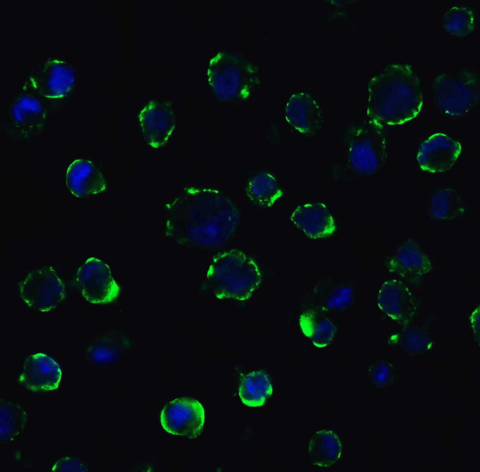
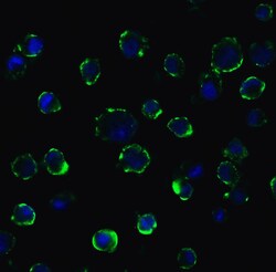
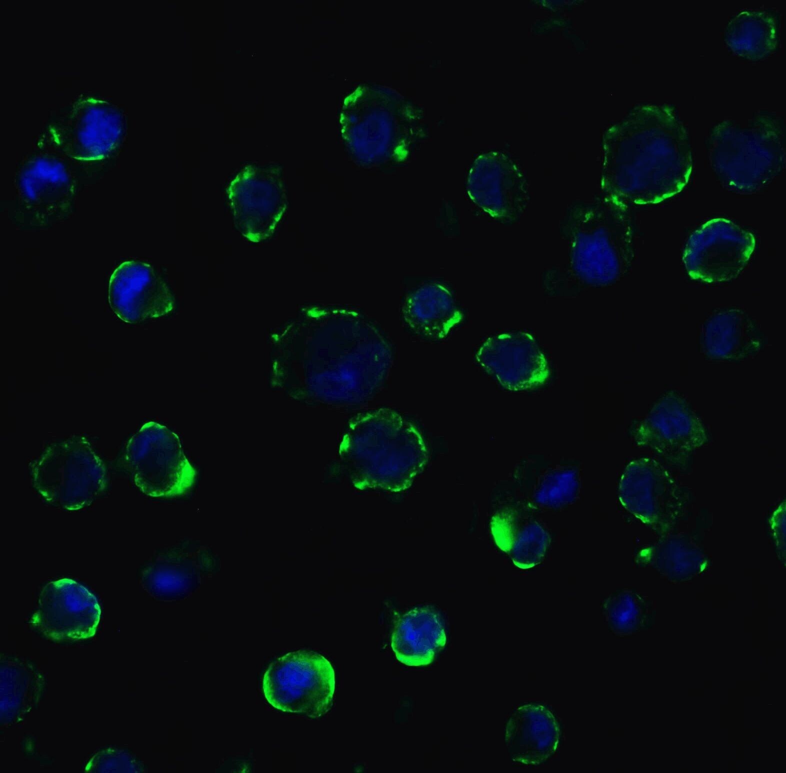
- Immunofluorescent analysis of 4% paraformaldehyde-fixed Caco2 cells labeling ACE2 with ACE2 Polyclonal Antibody (Product # PA5-20046) at 5 µg/mL, followed by goat anti-rabbit IgG secondary antibody at 1:500 dilution (green) and DAPI staining (blue). Image showing membrane staining on Caco2 cells.
- Submitted by
- Invitrogen Antibodies (provider)
- Main image

- Experimental details
- Immunofluorescent analysis of 4% paraformaldehyde-fixed Caco2 cells labeling ACE2 with ACE2 Polyclonal Antibody (Product # PA5-20046) at 5 µg/mL, followed by goat anti-rabbit IgG secondary antibody at 1:500 dilution (green) and DAPI staining (blue). Image showing membrane staining on Caco2 cells.

- Submitted by
- Invitrogen Antibodies (provider)
- Main image
- Experimental details
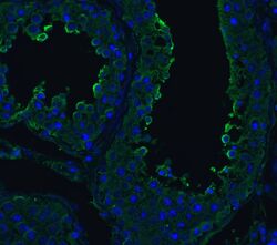
- Immunofluorescent analysis of 4% paraformaldehyde-fixed human kidney cells labeling ACE2 with ACE2 Polyclonal Antibody (Product # PA5-20046) at 20 µg/mL, followed by goat anti-rabbit IgG secondary antibody at 1:500 dilution (green).
- Submitted by
- Invitrogen Antibodies (provider)
- Main image
- Experimental details
- Immunofluorescent analysis of 4% paraformaldehyde-fixed Caco2 cells labeling ACE2 with ACE2 Polyclonal Antibody (Product # PA5-20046) at 5 µg/mL, followed by goat anti-rabbit IgG secondary antibody at 1:500 dilution (green) and DAPI staining (blue). Image showing membrane staining on Caco2 cells.
- Submitted by
- Invitrogen Antibodies (provider)
- Main image

- Experimental details
- Immunofluorescent analysis of 4% paraformaldehyde-fixed human kidney cells labeling ACE2 with ACE2 Polyclonal Antibody (Product # PA5-20046) at 20 µg/mL, followed by goat anti-rabbit IgG secondary antibody at 1:500 dilution (green).

- Submitted by
- Invitrogen Antibodies (provider)
- Main image
- Experimental details
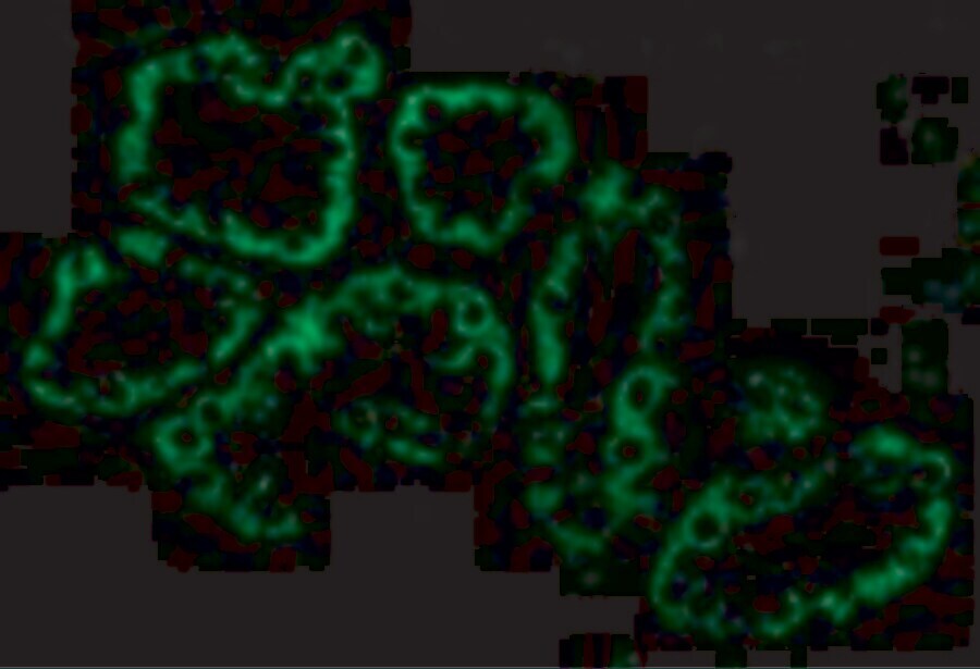
- Immunofluorescence analysis of ACE2 Polyclonal Antibody was performed in sections of H9 derived Lung organoids derived on Day 50. The organoids were fixed with 4% PFA for 1 hour at room temperature, followed by incubation in 30% sucrose solution overnight at 4°C. The organoids were then embedded in OCT and cryosectioned at 10 µm, permeabilized with 0.1% Triton™ X-100 for 10 minutes and blocked with 2% BSA for 1 hour at room temperature. The cells were labeled with ACE2 Polyclonal Antibody (Product # PA5-20046, 1:500) in 0.1% BSA and incubated for 3 hours at room temperature and then labeled with Goat anti-Rabbit IgG (H+L) Superclonal™ Secondary Antibody, Alexa Fluor™ 488 (Product # A27034, 1:2,000) for 45 minutes at room temperature. The sections were stained with DAPI (Product # D1306) and F-actin (Product # R415). The images were captured on EVOS™ M7000 Imaging System (Product # AMF7000) at 20X magnification.
Supportive validation
- Submitted by
- Invitrogen Antibodies (provider)
- Main image
- Experimental details
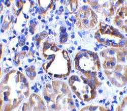
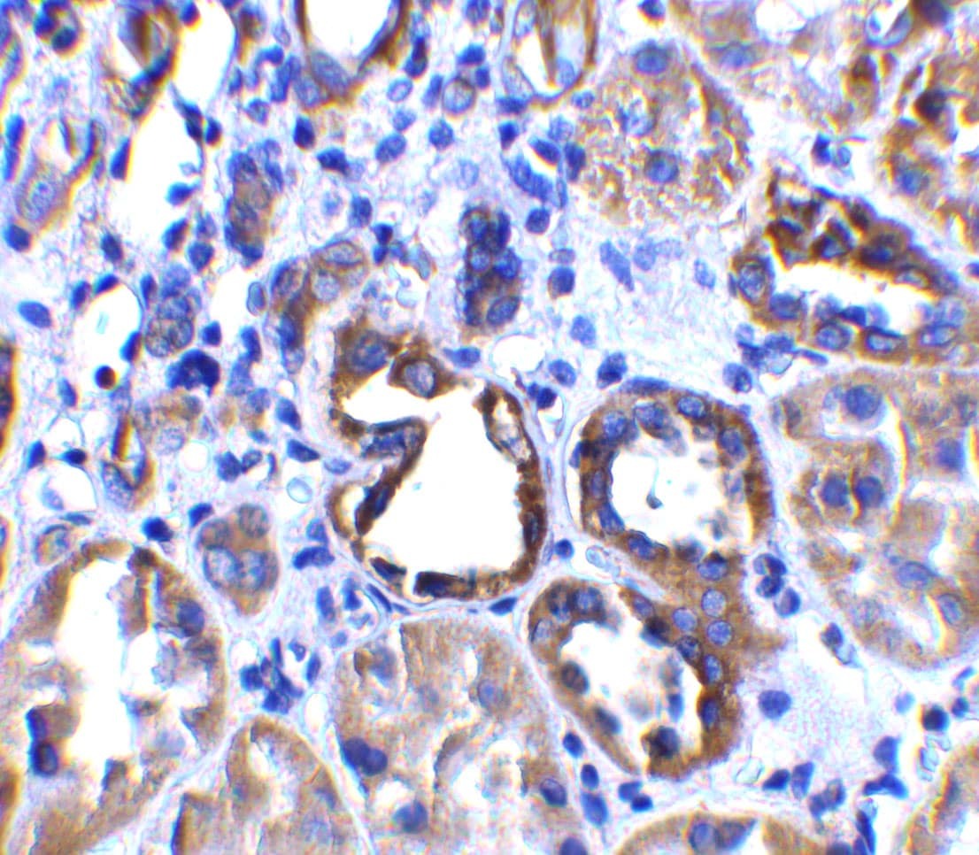
- Immunohistochemical analysis of paraffin-embedded human kidney tissue using ACE2 Polyclonal Antibody (Product # PA5-20046) at 2 µg/mL. Tissue was fixed with formaldehyde and blocked with 0.1 serum for 1 h at RT; antigen retrieval was by heat mediation with a citrate buffer (pH6). Samples were incubated with primary antibody overnight at 4 ˚C. A goat anti-rabbit IgG H&L (HRP) at 1/250 was used as secondary. Counter stained with Hematoxylin.
- Submitted by
- Invitrogen Antibodies (provider)
- Main image
- Experimental details
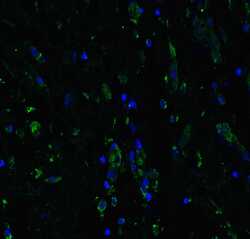
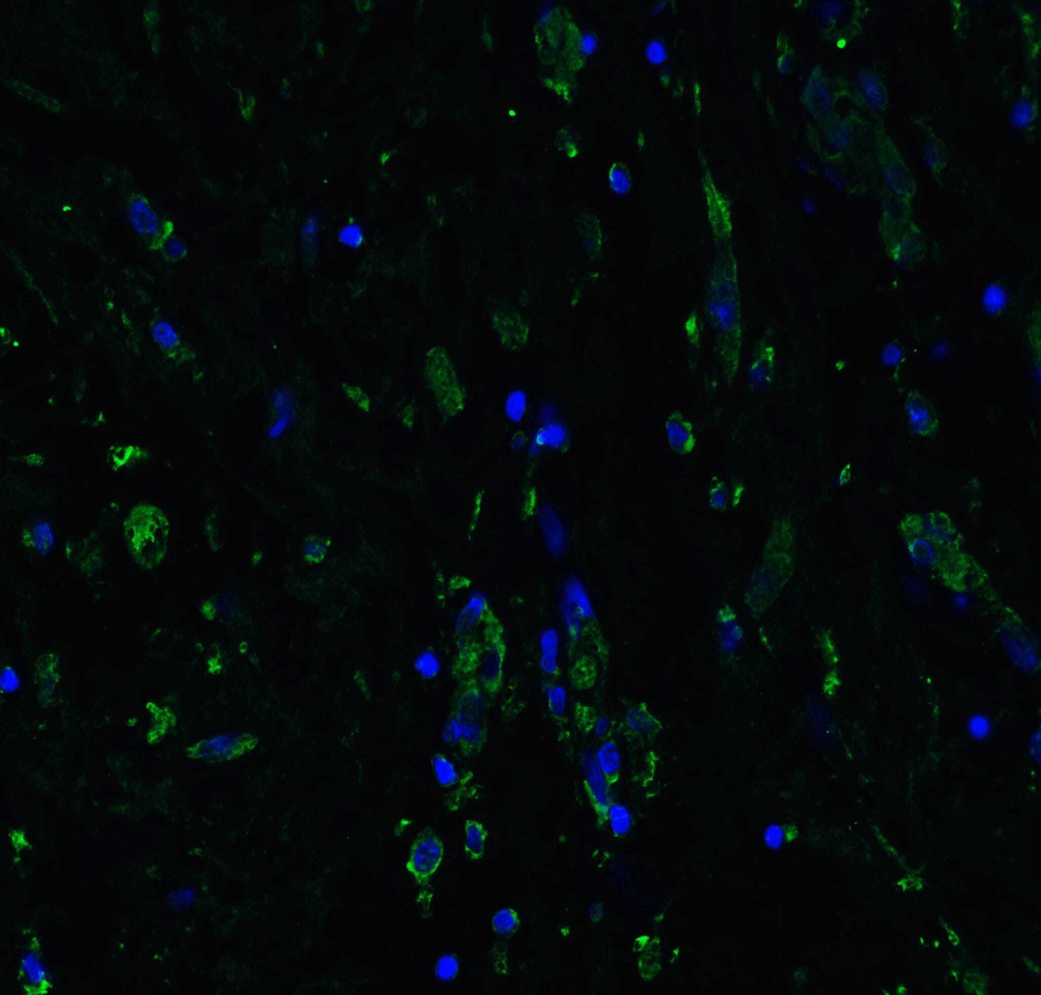
- Immunofluorescent analysis of 4% paraformaldehyde-fixed human testis tissue labeling ACE-2 with ACE2 Polyclonal Antibody (Product # PA5-20046) at 20 µg/mL, followed by goat anti-rabbit IgG secondary antibody at 1:500 dilution (green) and DAPI staining (blue).
- Submitted by
- Invitrogen Antibodies (provider)
- Main image
- Experimental details
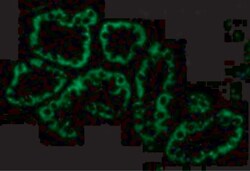
- Immunofluorescent analysis of 4% paraformaldehyde-fixed rat lung tissue labeling ACE-2 with ACE2 Polyclonal Antibody (Product # PA5-20046) at 20 µg/mL, followed by goat anti-rabbit IgG secondary antibody at 1:500 dilution (green) and DAPI staining (blue).
- Submitted by
- Invitrogen Antibodies (provider)
- Main image
- Experimental details
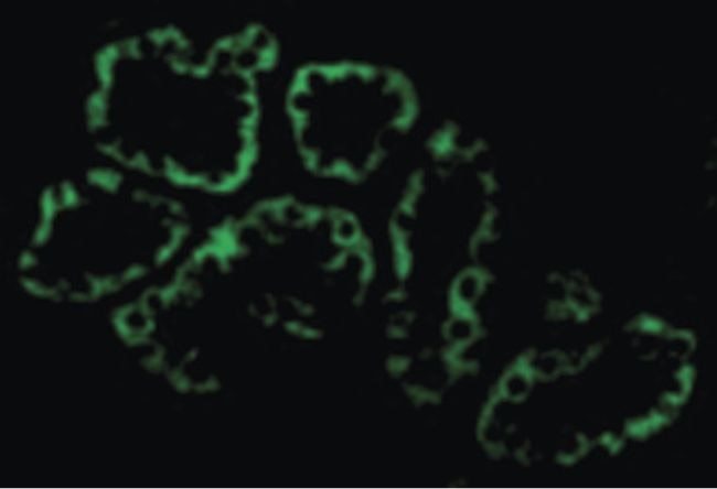
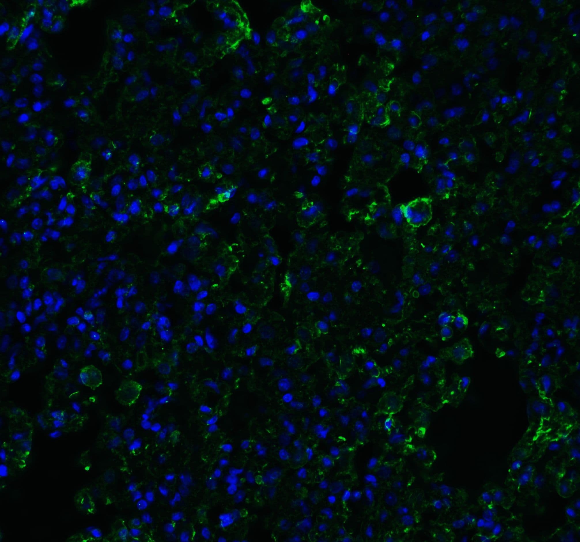
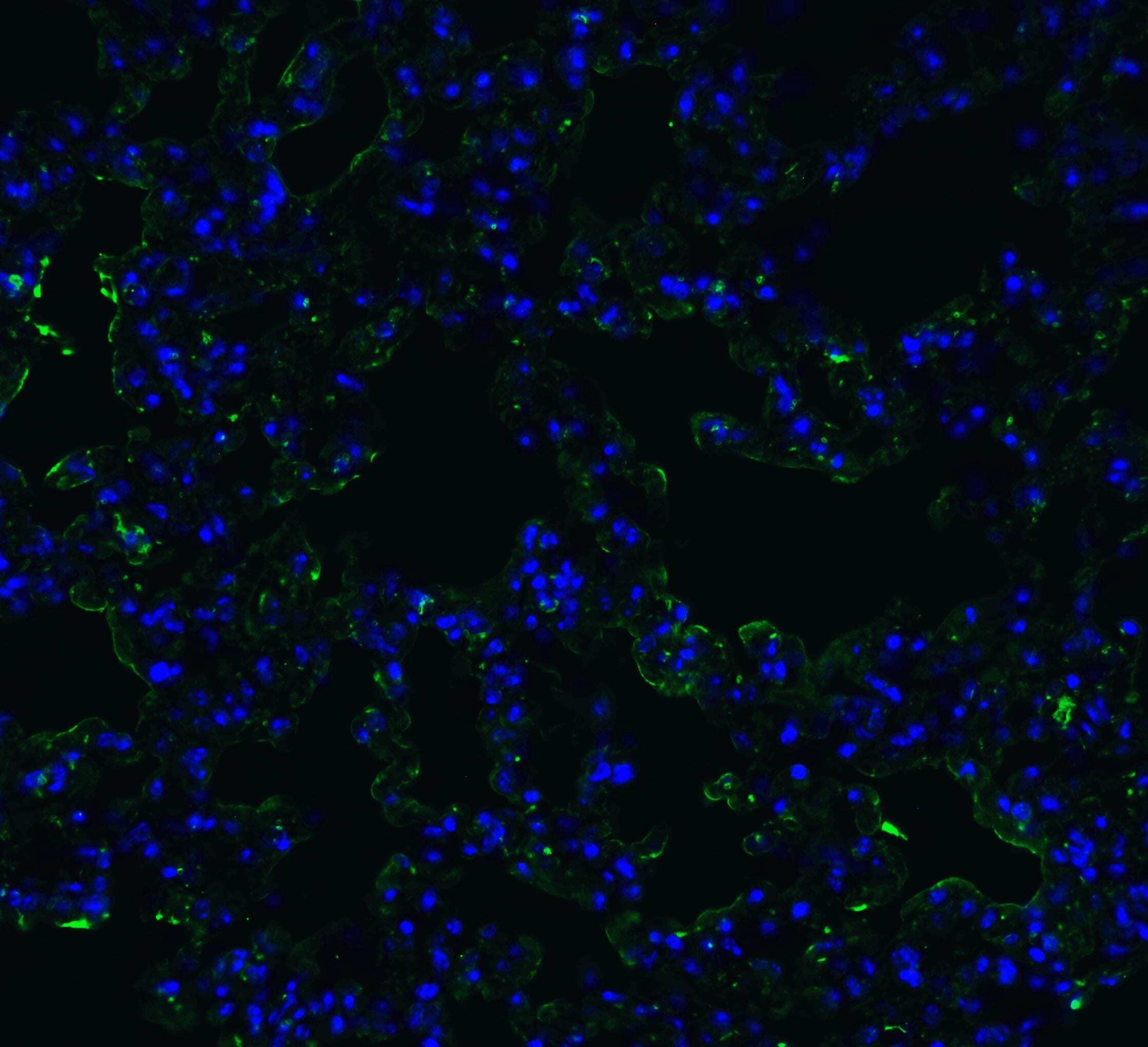
- Immunofluorescent analysis of 4% paraformaldehyde-fixed mouse lung tissue labeling ACE-2 with ACE2 Polyclonal Antibody (Product # PA5-20046) at 20 µg/mL, followed by goat anti-rabbit IgG secondary antibody at 1:500 dilution (green) and DAPI staining (blue).
- Submitted by
- Invitrogen Antibodies (provider)
- Main image
- Experimental details
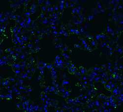
- Immunofluorescent analysis of 4% paraformaldehyde-fixed human lung tissue labeling ACE-2 with ACE2 Polyclonal Antibody (Product # PA5-20046) at 20 µg/mL, followed by goat anti-rabbit IgG secondary antibody at 1:500 dilution (green) and DAPI staining (blue).
Supportive validation
- Submitted by
- Invitrogen Antibodies (provider)
- Main image

- Experimental details
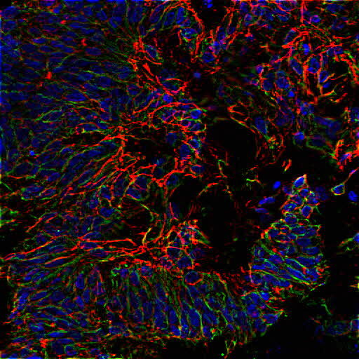
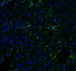
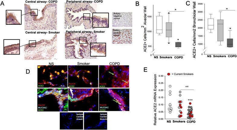
- Fig. 1 ACE2 expression in bronchial and alveolar epithelium from COPD patients, smoker and never-smoker (NS) controls. The number of ACE2+ cells in the central airway bronchial epithelium was similar between patients with chronic obstructive pulmonary disease (COPD), smokers without COPD and NS controls. In A , representative IHC for ACE2 images of central airways of a COPD patient (upper panel) and a smoker without COPD. The insets show details of the ciliated bronchial epithelium. The number of ACE2+ cells in the alveolar epithelium ( B ) and peripheral airway epithelium ( C ), normalized for length of the alveolar wall or basement membrane, respectively, was lower in patients with COPD versus smokers without COPD and NS controls. In D , triple immunofluorescence representative images of alveolar (upper panels) and bronchiolar epithelium (lower panels) from a COPD patient, a smoker without COPD, and a never-smoker (NS) where ACE2 staining is identified by green fluorochrome, the epithelium is identified by red fluorochrome, and the color yellow is obtained by merging the two fluorochromes. In E , the levels of ACE2 mRNA from peripheral lung samples were decreased between patients with chronic obstructive pulmonary disease (COPD) versus both smoker without COPD and NS controls. The red circles indicate the current smokers among the smoker controls and COPD patients
- Submitted by
- Invitrogen Antibodies (provider)
- Main image
- Experimental details
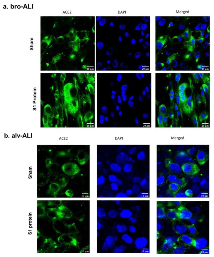
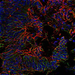
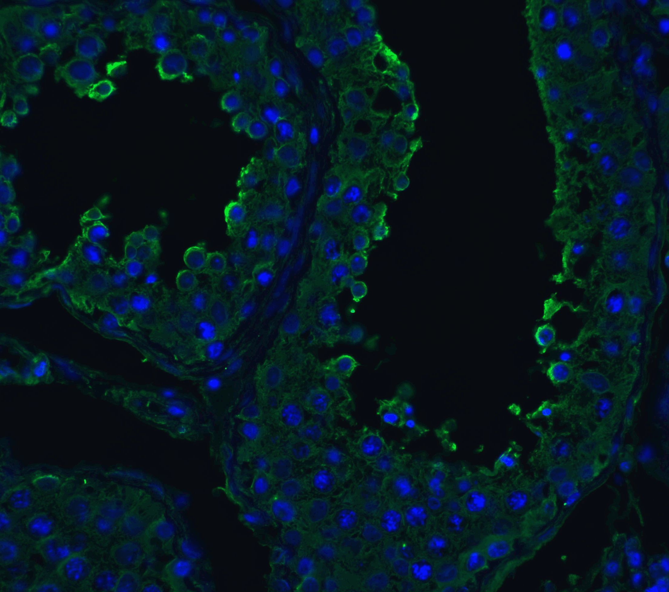
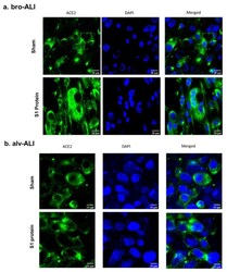
- Figure 3 Confocal microscopy of angiotensin converting enzyme 2 (ACE2) expression in the bronchial (bro-ALI: ( a ) and alveolar (alv-ALI: ( b ) mucosa model developed at air-liquid interface. Both bro-ALI and alv-ALI were exposed to 10 nM recombinant SARS-CoV-2 spike glycoprotein S1 (S1 protein) for 6 h and compared to the sham (representative picture of three independent observations). Scale bar bro-ALI: 20 um; scale bar alv-ALI: 10 um.