 Explore
 Validate
 Learn
Learn Flow cytometry
Antibody data
- Antibody Data
- Antigen structure
- References [2]
- Comments [0]
- Validations
- Flow cytometry [1]
Submit
Validation data
Reference
Comment
Report error
- Product number
- FAB946F - Provider product page

- Provider
- R&D Systems
- Product name
- Mouse ADAM10 Ectodomain Fluorescein-conjugated Antibody
- Antibody type
- Monoclonal
- Description
- Protein A or G purified from hybridoma culture supernatant. Detects mouse ADAM10 in direct ELISAs and Western blots. In direct ELISAs, approximately 25% cross-reactivity with recombinant human (rh) rhADAM10 is observed and no cross-reactivity with rhADAM8, recombinant mouse (rm) ADAM9, rmADAM15, rhBACE, or rhTACE is observed.
- Reactivity
- Mouse
- Host
- Rat
- Antigen sequence
O35598- Isotype
- IgG
- Antibody clone number
- 139712
- Vial size
- 100 Tests
- Storage
- Protect from light. Do not freeze. 12 months from date of receipt, 2 to 8 °C as supplied.
Submitted references Adrenergic regulation of IgE involves modulation of CD23 and ADAM10 expression on exosomes.
ADAM10 overexpression shifts lympho- and myelopoiesis by dysregulating site 2/site 3 cleavage products of Notch.
Padro CJ, Shawler TM, Gormley MG, Sanders VM
Journal of immunology (Baltimore, Md. : 1950) 2013 Dec 1;191(11):5383-97
Journal of immunology (Baltimore, Md. : 1950) 2013 Dec 1;191(11):5383-97
ADAM10 overexpression shifts lympho- and myelopoiesis by dysregulating site 2/site 3 cleavage products of Notch.
Gibb DR, Saleem SJ, Kang DJ, Subler MA, Conrad DH
Journal of immunology (Baltimore, Md. : 1950) 2011 Apr 1;186(7):4244-52
Journal of immunology (Baltimore, Md. : 1950) 2011 Apr 1;186(7):4244-52
No comments: Submit comment
Supportive validation
- Submitted by
- R&D Systems (provider)
- Main image
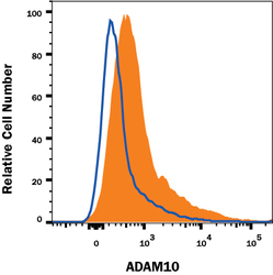
- Experimental details
- Detection of ADAM10 in Mouse Splenocytes by Flow Cytometry. Mouse splenocytes were stained with Rat Anti-Mouse ADAM10 Ectodomain Fluorescein-conjugated Monoclonal Antibody (Catalog # FAB946F, filled histogram) or isotype control antibody (Catalog # IC006F, open histogram). View our protocol for Staining Membrane-associated Proteins.
