 Explore
 Validate
 Learn
Learn Western blot
 Immunohistochemistry
 Flow cytometry
Antibody data
- Antibody Data
- Antigen structure
- References [9]
- Comments [0]
- Validations
- Flow cytometry [1]
- Other assay [1]
Submit
Validation data
Reference
Comment
Report error
- Product number
- PA1-973 - Provider product page

- Provider
- Invitrogen Antibodies
- Product name
- PSMD1 Polyclonal Antibody
- Antibody type
- Polyclonal
- Antigen
- Synthetic peptide
- Description
- PA1-973 detects proteasome 19S subunit S1 from from human cells. PA1-973 has been successfully used in Western blot and immunohistochemistry procedures. By Western blot, this antibody detects an ~105 kDa protein representing proteasome 19S subunit S1 from HeLa cell extract. PA1-973 immunizing peptide corresponds to amino acid residues 929-953 from human proteasome 19S subunit S1 protein.
- Reactivity
- Human, Mouse, Rat, Bovine
- Host
- Rabbit
- Isotype
- IgG
- Vial size
- 100 μg
- Concentration
- 1 mg/mL
- Storage
- -20°C, Avoid Freeze/Thaw Cycles
Submitted references Inactive USP14 and inactive UCHL5 cause accumulation of distinct ubiquitinated proteins in mammalian cells.
Impairments in age-dependent ubiquitin proteostasis and structural integrity of selective neurons by uncoupling Ran GTPase from the Ran-binding domain 3 of Ranbp2 and identification of novel mitochondrial isoforms of ubiquitin-conjugating enzyme E2I (ubc9) and Ranbp2.
Differential loss of prolyl isomerase or chaperone activity of Ran-binding protein 2 (Ranbp2) unveils distinct physiological roles of its cyclophilin domain in proteostasis.
Glucosamine induces cell death via proteasome inhibition in human ALVA41 prostate cancer cell.
Persistent hijacking of brain proteasomes in HIV-associated dementia.
The cyclophilin-like domain of Ran-binding protein-2 modulates selectively the activity of the ubiquitin-proteasome system and protein biogenesis.
Reduced ubiquitin C-terminal hydrolase-1 expression levels in dementia with Lewy bodies.
Neuropathology and pathogenesis of encephalitis following amyloid-beta immunization in Alzheimer's disease.
Neuropathology and pathogenesis of encephalitis following amyloid-beta immunization in Alzheimer's disease.
Chadchankar J, Korboukh V, Conway LC, Wobst HJ, Walker CA, Doig P, Jacobsen SJ, Brandon NJ, Moss SJ, Wang Q
PloS one 2019;14(11):e0225145
PloS one 2019;14(11):e0225145
Impairments in age-dependent ubiquitin proteostasis and structural integrity of selective neurons by uncoupling Ran GTPase from the Ran-binding domain 3 of Ranbp2 and identification of novel mitochondrial isoforms of ubiquitin-conjugating enzyme E2I (ubc9) and Ranbp2.
Patil H, Yoon D, Bhowmick R, Cai Y, Cho KI, Ferreira PA
Small GTPases 2019 Mar;10(2):146-161
Small GTPases 2019 Mar;10(2):146-161
Differential loss of prolyl isomerase or chaperone activity of Ran-binding protein 2 (Ranbp2) unveils distinct physiological roles of its cyclophilin domain in proteostasis.
Cho KI, Patil H, Senda E, Wang J, Yi H, Qiu S, Yoon D, Yu M, Orry A, Peachey NS, Ferreira PA
The Journal of biological chemistry 2014 Feb 21;289(8):4600-25
The Journal of biological chemistry 2014 Feb 21;289(8):4600-25
Glucosamine induces cell death via proteasome inhibition in human ALVA41 prostate cancer cell.
Liu BQ, Meng X, Li C, Gao YY, Li N, Niu XF, Guan Y, Wang HQ
Experimental & molecular medicine 2011 Sep 30;43(9):487-93
Experimental & molecular medicine 2011 Sep 30;43(9):487-93
Persistent hijacking of brain proteasomes in HIV-associated dementia.
Nguyen TP, Soukup VM, Gelman BB
The American journal of pathology 2010 Feb;176(2):893-902
The American journal of pathology 2010 Feb;176(2):893-902
The cyclophilin-like domain of Ran-binding protein-2 modulates selectively the activity of the ubiquitin-proteasome system and protein biogenesis.
Yi H, Friedman JL, Ferreira PA
The Journal of biological chemistry 2007 Nov 30;282(48):34770-8
The Journal of biological chemistry 2007 Nov 30;282(48):34770-8
Reduced ubiquitin C-terminal hydrolase-1 expression levels in dementia with Lewy bodies.
Barrachina M, Castaño E, Dalfó E, Maes T, Buesa C, Ferrer I
Neurobiology of disease 2006 May;22(2):265-73
Neurobiology of disease 2006 May;22(2):265-73
Neuropathology and pathogenesis of encephalitis following amyloid-beta immunization in Alzheimer's disease.
Ferrer I, Boada Rovira M, Sánchez Guerra ML, Rey MJ, Costa-Jussá F
Brain pathology (Zurich, Switzerland) 2004 Jan;14(1):11-20
Brain pathology (Zurich, Switzerland) 2004 Jan;14(1):11-20
Neuropathology and pathogenesis of encephalitis following amyloid-beta immunization in Alzheimer's disease.
Ferrer I, Boada Rovira M, Sánchez Guerra ML, Rey MJ, Costa-Jussá F
Brain pathology (Zurich, Switzerland) 2004 Jan;14(1):11-20
Brain pathology (Zurich, Switzerland) 2004 Jan;14(1):11-20
No comments: Submit comment
Supportive validation
- Submitted by
- Invitrogen Antibodies (provider)
- Main image
- Experimental details
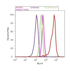
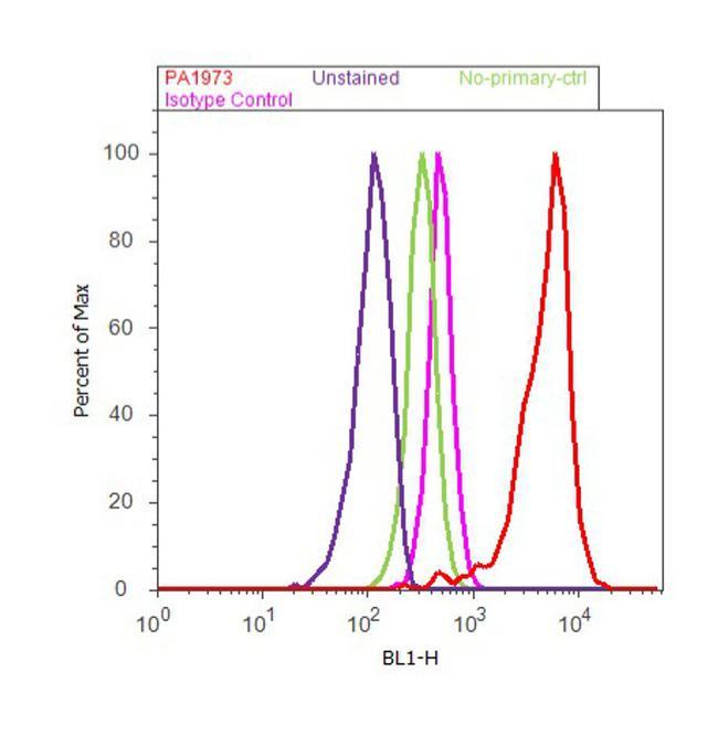
- Flow cytometry analysis of PSMD1 was done on MCF7 cells. Cells were fixed with 70% ethanol for 10 minutes, permeabilized with 0.25% Triton™ X-100 for 20 minutes, and blocked with 5% BSA for 30 minutes at room temperature. Cells were labeled with PSMD1 Rabbit Polyclonal Antibody (PA1973, red histogram) or with rabbit isotype control (pink histogram) at 3-5 ug/million cells in 2.5% BSA. After incubation at room temperature for 2 hours, the cells were labeled with Alexa Fluor® 488 Goat Anti-Rabbit Secondary Antibody (A11008) at a dilution of 1:400 for 30 minutes at room temperature. The representative 10,000 cells were acquired and analyzed for each sample using an Attune® Acoustic Focusing Cytometer. The purple histogram represents unstained control cells and the green histogram represents no-primary-antibody control..
Supportive validation
- Submitted by
- Invitrogen Antibodies (provider)
- Main image

- Experimental details
- NULL
