 Explore
 Validate
 Learn
Learn Western blot
 Immunocytochemistry
Antibody data
- Antibody Data
- Antigen structure
- References [2]
- Comments [0]
- Validations
- Immunocytochemistry [2]
Submit
Validation data
Reference
Comment
Report error
- Product number
- PA1-726 - Provider product page

- Provider
- Invitrogen Antibodies
- Product name
- PDE6D Polyclonal Antibody
- Antibody type
- Polyclonal
- Antigen
- Synthetic peptide
- Description
- PA1-726 detects PrBP/delta from bovine, human and sheep samples. PA1-726 has been successfully used in Western blot, ICC/IF and immunohistochemistry procedures. By Western blot, this antibody detects a 17 kDa protein representing PrBP/delta from bovine and sheep retinal/optic nerve extracts as well as HeLa cell lysate. Immunohistochemical staining of PrBP/delta in bovine retinal cross-sections using PA1-726 results in staining of retinal outer segments. PA1-726 immunizing peptides correspond to amino acid residues 1-16 from mouse and human PrBP/delta. This peptide (Cat. # PEP-203) is available for use in neutralization and control experiments.
- Reactivity
- Human, Bovine
- Host
- Rabbit
- Isotype
- IgG
- Vial size
- 100 μg
- Concentration
- 1 mg/mL
- Storage
- -20°C, Avoid Freeze/Thaw Cycles
Submitted references Structural and functional protein network analyses predict novel signaling functions for rhodopsin.
Mammalian sperm phosphodiesterases and their involvement in receptor-mediated cell signaling important for capacitation.
Kiel C, Vogt A, Campagna A, Chatr-aryamontri A, Swiatek-de Lange M, Beer M, Bolz S, Mack AF, Kinkl N, Cesareni G, Serrano L, Ueffing M
Molecular systems biology 2011 Nov 22;7:551
Molecular systems biology 2011 Nov 22;7:551
Mammalian sperm phosphodiesterases and their involvement in receptor-mediated cell signaling important for capacitation.
Baxendale RW, Fraser LR
Molecular reproduction and development 2005 Aug;71(4):495-508
Molecular reproduction and development 2005 Aug;71(4):495-508
No comments: Submit comment
Supportive validation
- Submitted by
- Invitrogen Antibodies (provider)
- Main image
- Experimental details
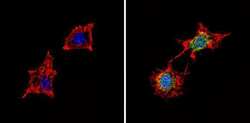
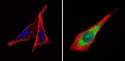
- Immunofluorescent analysis of PDE6 delta (green) showing staining in the cytoplasm and nucleus of B-3 cells (right) compared to a negative control without primary antibody (left). Formalin-fixed cells were permeabilized with 0.1% Triton X-100 in TBS for 5-10 minutes and blocked with 3% BSA-PBS for 30 minutes at room temperature. Cells were probed with a PDE6 delta polyclonal antibody (Product # PA1-726) in 3% BSA-PBS at a dilution of 1:100 and incubated overnight at 4 ºC in a humidified chamber. Cells were washed with PBST and incubated with a DyLight-conjugated secondary antibody in PBS at room temperature in the dark. F-actin (red) was stained with a fluorescent red phalloidin and nuclei (blue) were stained with Hoechst or DAPI. Images were taken at a magnification of 60x.
- Submitted by
- Invitrogen Antibodies (provider)
- Main image
- Experimental details
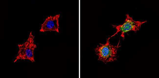
- Immunofluorescent analysis of PDE6 delta (green) showing staining in the cytoplasm and nucleus of B-3 cells (right) compared to a negative control without primary antibody (left). Formalin-fixed cells were permeabilized with 0.1% Triton X-100 in TBS for 5-10 minutes and blocked with 3% BSA-PBS for 30 minutes at room temperature. Cells were probed with a PDE6 delta polyclonal antibody (Product # PA1-726) in 3% BSA-PBS at a dilution of 1:100 and incubated overnight at 4 ºC in a humidified chamber. Cells were washed with PBST and incubated with a DyLight-conjugated secondary antibody in PBS at room temperature in the dark. F-actin (red) was stained with a fluorescent red phalloidin and nuclei (blue) were stained with Hoechst or DAPI. Images were taken at a magnification of 60x.
