Explore
 Validate
 Learn
Learn Immunocytochemistry
 Immunohistochemistry
Antibody data
- Antibody Data
- Antigen structure
- References [1]
- Comments [0]
- Validations
- Immunocytochemistry [1]
Submit
Validation data
Reference
Comment
Report error
- Product number
- AMAb91164 - Provider product page

- Provider
- Atlas Antibodies
- Proper citation
- Atlas Antibodies Cat#AMAb91164, RRID:AB_2665828
- Product name
- Anti-CEP350
- Antibody type
- Monoclonal
- Description
- Monoclonal Antibody against Human CEP350, Clone ID: CL3423, Gene description: centrosomal protein 350kDa, Alternative Gene Names: CAP350, KIAA0480, Validated applications: ICC, IHC, Uniprot ID: Q5VT06, Storage: Store at +4°C for short term storage. Long time storage is recommended at -20°C.
- Reactivity
- Human
- Host
- Mouse
- Conjugate
- Unconjugated
- Isotype
- IgG
- Antibody clone number
- CL3423
- Vial size
- 100 µl
- Concentration
- 0.5 mg/ml
- Storage
- Store at +4°C for short term storage. Long time storage is recommended at -20°C.
- Handling
- The antibody solution should be gently mixed before use.
Submitted references Trip13 Depletion in Liver Cancer Induces a Lipogenic Response Contributing to Plin2‐Dependent Mitotic Cell Death
Rios Garcia M, Meissburger B, Chan J, de Guia R, Mattijssen F, Roessler S, Birkenfeld A, Raschzok N, Riols F, Tokarz J, Giroud M, Gil Lozano M, Hartleben G, Nawroth P, Haid M, López M, Herzig S, Berriel Diaz M
Advanced Science 2022;9(29)
Advanced Science 2022;9(29)
No comments: Submit comment
Supportive validation
- Submitted by
- Atlas Antibodies (provider)
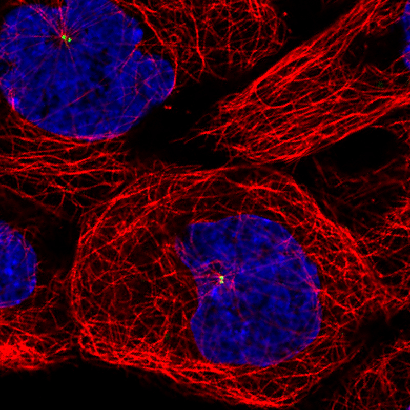
- Main image

- Experimental details
- Immunofluorescence staining of A431 cells using the anti-CEP350 monoclonal antibody, showing specific staining of the centrosome in green. Microtubule- and nuclear probes are visualized in red and blue, respectively (where available).
- Sample type
- Human