 Explore
 Validate
 Learn
Learn Western blot
Antibody data
- Antibody Data
- Antigen structure
- References [0]
- Comments [0]
- Validations
- Western blot [3]
- Immunocytochemistry [2]
- Immunoprecipitation [1]
- Other assay [1]
Submit
Validation data
Reference
Comment
Report error
- Product number
- 710855 - Provider product page

- Provider
- Invitrogen Antibodies
- Product name
- Syntaxin 6 Recombinant Superclonal™ Antibody (8HCLC)
- Antibody type
- Other
- Antigen
- Synthetic peptide
- Description
- This antibody is predicted to react with Monkey, Pig, Bovine, Rat and Mouse. Recombinant rabbit Superclonal™ antibodies are unique offerings from Thermo Fisher Scientific. They are comprised of a selection of multiple different recombinant monoclonal antibodies, providing the best of both worlds - the sensitivity of polyclonal antibodies with the specificity of monoclonal antibodies - all delivered with the consistency only found in a recombinant antibody. While functionally the same as a polyclonal antibody - recognizing multiple epitope sites on the target and producing higher detection sensitivity for low abundance targets - a recombinant rabbit Superclonal™ antibody has a known mixture of light and heavy chains. The exact population can be produced in every lot, circumventing the biological variability typically associated with polyclonal antibody production. Note: Formerly called “Recombinant polyclonal antibody”, this product is now rebranded as “Recombinant Superclonal™ antibody”. The physical product and the performance remain unchanged.
- Reactivity
- Human
- Host
- Rabbit
- Isotype
- IgG
- Antibody clone number
- 8HCLC
- Vial size
- 100 μg
- Concentration
- 0.5 mg/mL
- Storage
- Store at 4°C short term. For long term storage, store at -20°C, avoiding freeze/thaw cycles.
No comments: Submit comment
Supportive validation
- Submitted by
- Invitrogen Antibodies (provider)
- Main image
- Experimental details
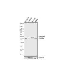
- Western blot analysis was performed on whole cell extracts (30 µg lysate) of SH-SY5Y (Lane1), U-87MG (Lane 2), SK-N-SH (Lane 3) and MDCK (Lane 4). The blots were probed with Anti-Syntaxin 6 Recombinant Rabbit Superclonal™ Antibody (Product # 710855, 0.5-1 µg/mL) and detected by chemiluminescence using Goat anti-Rabbit IgG (Heavy Chain) Superclonal™ Secondary Antibody, HRP conjugate (Product # A27036, 0.4 µg/mL, 1:2500 dilution). A clear 30 kDa band corresponding to Syntaxin 6 was observed across cell lines tested. Known quantity of protein samples were electrophoresed using Novex®NuPAGE®4-12% Bis-Tris gel (Product # NP0321BOX), XCell SureLock™ Electrophoresis System (Product # EI0002), and Novex® Sharp Pre-Stained Protein Standard (Product # LC5800). Resolved proteins were then transferred onto a nitrocellulose membrane with iBlot® Dry Blotting System (Product # IB21001). The membrane was probed with the relevant primary and secondary Antibody following blocking with 5% skimmed milk. Chemiluminescent detection was performed using Pierce™ ECL Western blotting Substrate (Product # 32106).

- Submitted by
- Invitrogen Antibodies (provider)
- Main image
- Experimental details
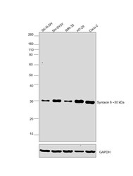
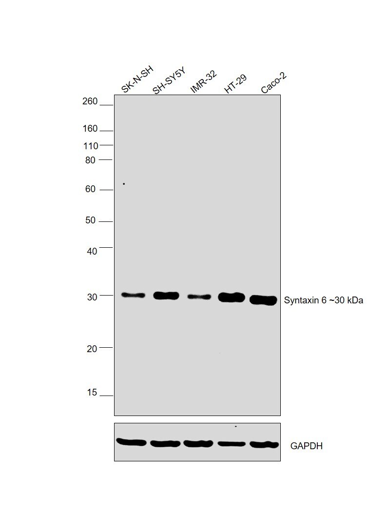
- Western blot was performed using Syntaxin 6 Recombinant Rabbit Superclonal™ Antibody (Product # 710855) and a 30kDa band corresponding to Syntaxin 6 was observed in the cell lines tested. Whole cell extracts (30 µg lysate) of SK-N-SH (Lane 1), SH-SY5Y (Lane 2), IMR-32 (Lane 3), HT-29 (Lane 4) and Caco-2 (Lane 5) were electrophoresed using NuPAGE™ 4-12% Bis-Tris Protein Gel (Product # NP0321BOX). Resolved proteins were then transferred onto a nitrocellulose membrane (Product # LC2001) by iBlot® 2 Dry Blotting System (Product # IB21001). The blot was probed with the primary antibody (1:1000 dilution) and detected by chemiluminescence with Goat anti-Rabbit IgG (Heavy Chain) Superclonal™ Recombinant Secondary Antibody, HRP (Product # A27036, 1:4000 dilution) using the iBright FL 1000 (Product # A32752). Chemiluminescent detection was performed using Novex® ECL Chemiluminescent Substrate Reagent Kit (Product # WP20005).
- Submitted by
- Invitrogen Antibodies (provider)
- Main image

- Experimental details
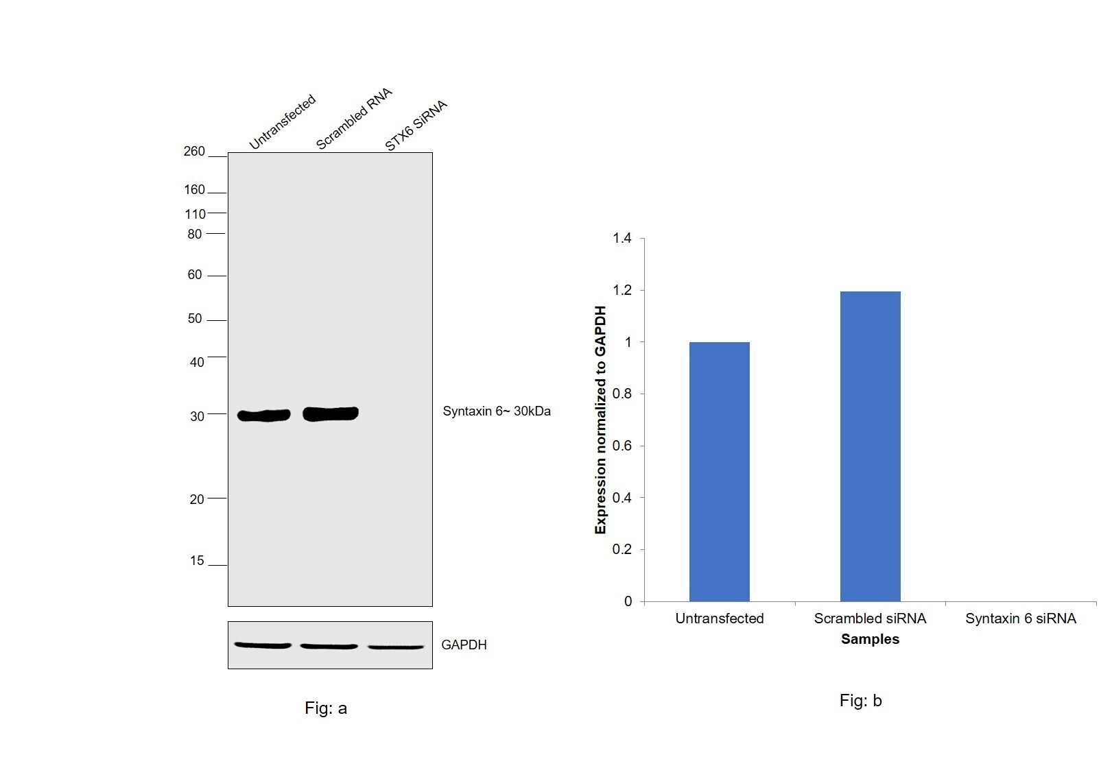
- Knockdown of Syntaxin 6 was achieved by transfecting SH-SY5Y with Syntaxin 6 specific siRNA (Silencer® select Product # s19959, s19958). Western blot analysis (Fig. a) was performed using modified whole cell extracts (1% SDS) from the Syntaxin 6 knockdown cells (lane 3), non-specific scrambled siRNA transfected cells (lane 2) and untransfected cells (lane 1). The blot was probed with Syntaxin 6 Recombinant Rabbit Superclonal™ Antibody (Product # 710855, 1:1,000 dilution) and Goat anti-Rabbit IgG (Heavy Chain) Superclonal™ Recombinant Secondary Antibody, HRP (Product # A27036, 0.25 µg/mL, 1:4,000 dilution). Densitometric analysis of this western blot is shown in histogram (Fig. b). Decrease in signal upon siRNA mediated knock down confirms that antibody is specific to Syntaxin 6.
Supportive validation
- Submitted by
- Invitrogen Antibodies (provider)
- Main image
- Experimental details
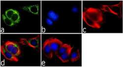
- Immunofluorescence was performed on fixed and permeabilized MCF-7 cells for detection of Syntaxin-6 using Anti-Syntaxin-6 Recombinant Rabbit Polyclonal Antibody (Product # 710855, 1 µg/mL) and labeled with Goat anti-Rabbit IgG (H+L) Superclonal™ Secondary Antibody, Alexa Fluor® 488 conjugate (Product # A27034, 1:2000). Panel a) shows representative cells that were stained for detection and localization of Syntaxin-6 protein (green), Panel b) is stained for nuclei (blue) using SlowFade® Gold Antifade Mountant with DAPI (Product # S36938,). Panel c) represents cytoskeletal F-actin staining using Alexa Fluor® 555 Rhodamine Phalloidin (Product # R415, 1:300). Panel d) is a composite image of Panels a, b and c clearly demonstrating cytoplasmic localization of Syntaxin-6. Panel e) represents control cells with no primary Antibody to assess background.

- Submitted by
- Invitrogen Antibodies (provider)
- Main image
- Experimental details
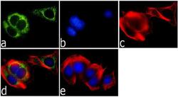
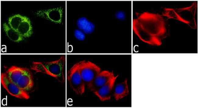
- Immunofluorescence was performed on fixed and permeabilized MCF-7 cells for detection of Syntaxin-6 using Anti-Syntaxin-6 Recombinant Rabbit Superclonal™ Antibody (Product # 710855, 1 µg/mL) and labeled with Goat anti-Rabbit IgG (Heavy Chain) Superclonal™ Secondary Antibody, Alexa Fluor® 488 conjugate (Product # A27034, 1:2000). Panel a) shows representative cells that were stained for detection and localization of Syntaxin-6 protein (green), Panel b) is stained for nuclei (blue) using SlowFade® Gold Antifade Mountant with DAPI (Product # S36938,). Panel c) represents cytoskeletal F-actin staining using Alexa Fluor® 555 Rhodamine Phalloidin (Product # R415, 1:300). Panel d) is a composite image of Panels a, b and c clearly demonstrating cytoplasmic localization of Syntaxin-6. Panel e) represents control cells with no primary Antibody to assess background.
Supportive validation
- Submitted by
- Invitrogen Antibodies (provider)
- Main image

- Experimental details
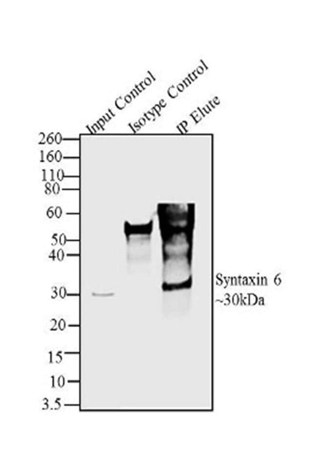
- Syntaxin 6 was immunoprecipitated using 2 µg of the Syntaxin 6 Recombinant Rabbit Superclonal™ Antibody (Product # 710855) from SK-N-SH cell extracts (Lane 3) using the Dynabeads® Protein A Immunoprecipitation Kit (Product # 10006D). Normal Rabbit IgG was used as a negative IP control (Lane 2). 10% input represents the cell extract used for immunoprecipitation (Lane 1). Western blot analysis was performed using Syntaxin 6 Recombinant Rabbit Superclonal™ Antibody (Product # 710855) and Goat anti-Rabbit IgG (Heavy Chain) Superclonal™ Secondary Antibody, HRP conjugate (Product # A27036, 0.4 µg/mL, 1:2500 dilution). Chemiluminescent detection was performed using Pierce™ ECL Western blotting Substrate (Product # 32106).
Supportive validation
- Submitted by
- Invitrogen Antibodies (provider)
- Main image

- Experimental details
- Syntaxin 6 was immunoprecipitated using 2 æg of the Syntaxin 6 Recombinant Rabbit Polyclonal Antibody (Product # 710855) from SK-N-SH cell extracts (Lane 3) using the Dynabeads® Protein A Immunoprecipitation Kit (Product # 10006D). Normal Rabbit IgG was used as a negative IP control (Lane 2). 10% input represents the cell extract used for immunoprecipitation (Lane 1). Western blot analysis was performed using Syntaxin 6 Recombinant Rabbit Polyclonal Antibody (Product # 710855) and Goat anti-Rabbit IgG (H+L) Superclonal™ Secondary Antibody, HRP conjugate (Product # A27036, 0.4 æg/mL, 1:2500 dilution). Chemiluminescent detection was performed using Pierce™ ECL Western blotting Substrate (Product # 32106).
