Explore
 Validate
 Learn
Learn Immunocytochemistry
Antibody data
- Antibody Data
- Antigen structure
- References [1]
- Comments [0]
- Validations
- Immunocytochemistry [1]
- Immunohistochemistry [1]
Submit
Validation data
Reference
Comment
Report error
- Product number
- HPA056959 - Provider product page

- Provider
- Atlas Antibodies
- Proper citation
- Atlas Antibodies Cat#HPA056959, RRID:AB_2683288
- Product name
- Anti-EFHD1
- Antibody type
- Polyclonal
- Description
- Polyclonal Antibody against Human EFHD1, Gene description: EF-hand domain family, member D1, Alternative Gene Names: FLJ13612, Validated applications: IHC, ICC, Uniprot ID: Q9BUP0, Storage: Store at +4°C for short term storage. Long time storage is recommended at -20°C.
- Reactivity
- Human
- Host
- Rabbit
- Conjugate
- Unconjugated
- Isotype
- IgG
- Vial size
- 100 µl
- Concentration
- 0.1 mg/ml
- Storage
- Store at +4°C for short term storage. Long time storage is recommended at -20°C.
- Handling
- The antibody solution should be gently mixed before use.
Submitted references Lineage-specific differences and regulatory networks governing human chondrocyte development
Pregizer S, Richard D, Venkatasubramanian D, Raftery R, Muthuirulan P, Liu Z, Capellini T, Craft A
eLife 2023;12
eLife 2023;12
No comments: Submit comment
Supportive validation
- Submitted by
- Atlas Antibodies (provider)
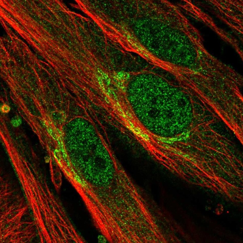
- Main image

- Experimental details
- Immunofluorescent staining of human cell line BJ shows localization to nucleoplasm & the Golgi apparatus.
- Sample type
- Human
Supportive validation
- Submitted by
- Atlas Antibodies (provider)
- Enhanced method
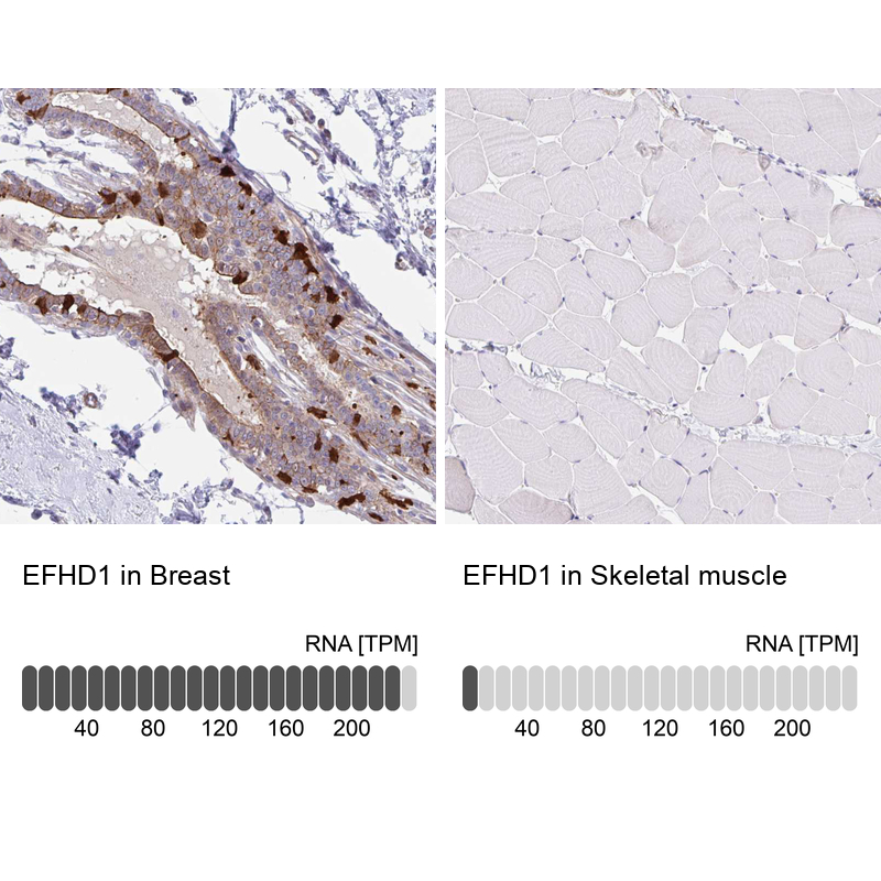
- Orthogonal validation
- Main image
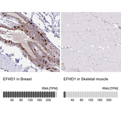
- Experimental details
- Immunohistochemistry analysis in human breast and skeletal muscle tissues using HPA056959 antibody. Corresponding EFHD1 RNA-seq data are presented for the same tissues.
- Sample type
- Human
- Protocol
- Protocol