Explore
 Validate
 Learn
Learn Western blot
 Immunocytochemistry
Antibody data
- Antibody Data
- Antigen structure
- References [1]
- Comments [0]
- Validations
- Immunocytochemistry [2]
- Immunohistochemistry [2]
- Flow cytometry [2]
- Other assay [1]
Submit
Validation data
Reference
Comment
Report error
- Product number
- PA5-35114 - Provider product page

- Provider
- Invitrogen Antibodies
- Product name
- NEU2 Polyclonal Antibody
- Antibody type
- Polyclonal
- Antigen
- Synthetic peptide
- Reactivity
- Human
- Host
- Rabbit
- Isotype
- IgG
- Vial size
- 400 μL
- Concentration
- 0.5 mg/mL
- Storage
- Store at 4°C short term. For long term storage, store at -20°C, avoiding freeze/thaw cycles.
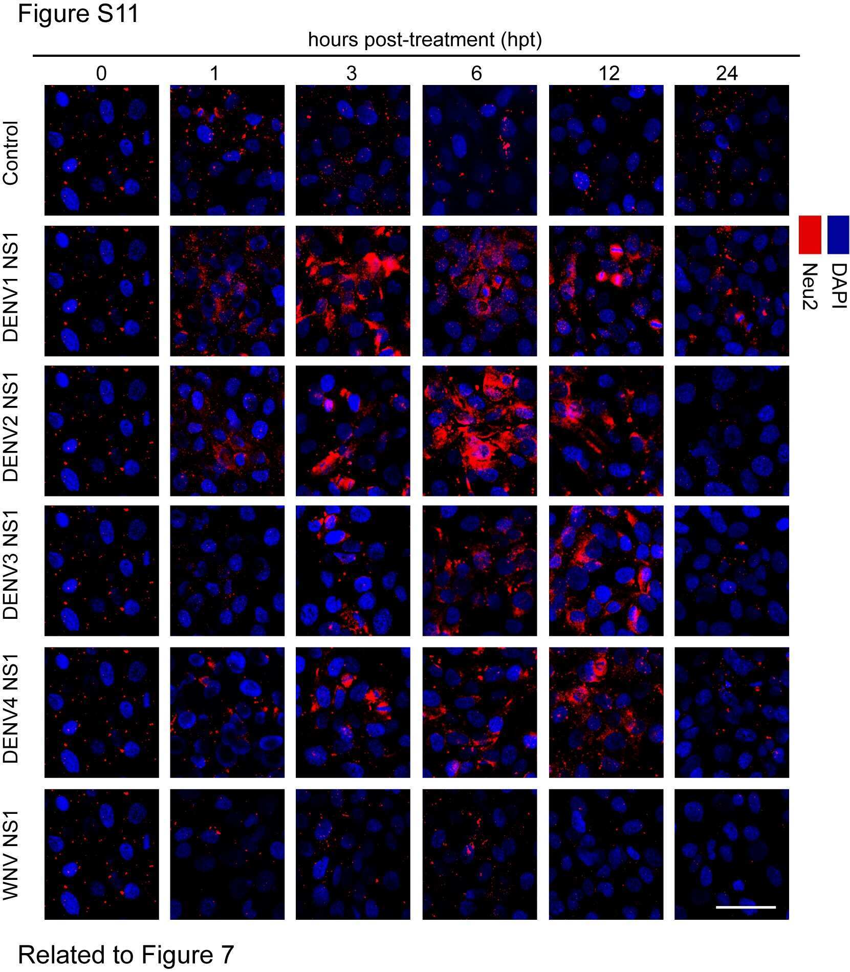
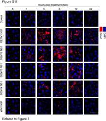
Submitted references Dengue Virus NS1 Disrupts the Endothelial Glycocalyx, Leading to Hyperpermeability.
Puerta-Guardo H, Glasner DR, Harris E
PLoS pathogens 2016 Jul;12(7):e1005738
PLoS pathogens 2016 Jul;12(7):e1005738
No comments: Submit comment
Supportive validation
- Submitted by
- Invitrogen Antibodies (provider)
- Main image

- Experimental details
- Immunofluorescent analysis of NEU2 in A549 cells using a NEU2 polyclonal antibody (Product # PA5-35114) followed by detection using a fluorescent conjugated secondary antibody (green). Nuclei were stained with Dapi (blue) and cytoplasmic actin was stained with a fluorescent phalloidin (red).

- Submitted by
- Invitrogen Antibodies (provider)
- Main image
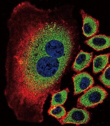
- Experimental details
- Immunocytochemistry analysis of NEU2 in A549 cells. Samples were incubated in NEU2 polyclonal antibody (Product # PA5-35114) followed by Alexa Fluor 488-conjugated goat anti-rabbit lgG (green). Actin filaments have been labeled with Alexa Fluor 555 phalloidin (red). DAPI was used to stain the cell nuclear (blue).

Supportive validation
- Submitted by
- Invitrogen Antibodies (provider)
- Main image

- Experimental details
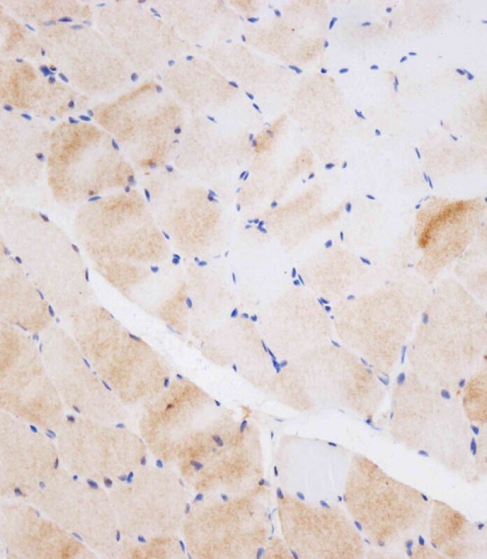
- Immunohistochemistry analysis of NEU2 in paraffin-embedded human skeletal muscle tissue. Samples were incubated with NEU2 polyclonal antibody (Product # PA5-35114) using a dilution of 1:250 for 15 min at room temperature followed by Leica Bond Polymer Refine Detection. Tissue was fixed with formaldehyde at room temperature. Heat induced epitope retrieval was performed by EDTA buffer (pH 9 0).
- Submitted by
- Invitrogen Antibodies (provider)
- Main image
- Experimental details
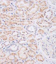
- Immunohistochemistry analysis of NEU2 in paraffin-embedded Human kidney tissue. Samples were incubated with NEU2 polyclonal antibody (Product # PA5-35114) using a dilution of 1:250 for 15 min at room temperature followed by Leica Bond Polymer Refine Detection. Tissue was fixed with formaldehyde at room temperature. Heat induced epitope retrieval was performed by EDTA buffer (pH 9 0).

Supportive validation
- Submitted by
- Invitrogen Antibodies (provider)
- Main image
- Experimental details
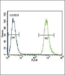
- Flow cytometry analysis of NEU2 in A549 cells (right) compared to a negative control (left) using a NEU2 polyclonal antibody (Product # PA5-35114) followed by detection using a FITC-conjugated goat-anti-rabbit secondary antibody.
- Submitted by
- Invitrogen Antibodies (provider)
- Main image
- Experimental details
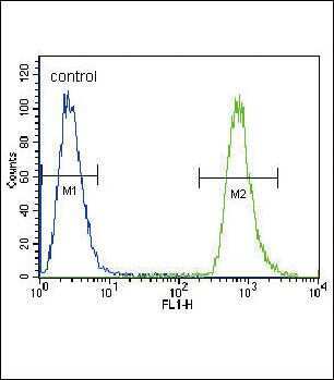
- Flow cytometry of NEU2 in A549 cells (right histogram). Samples were incubated with NEU2 polyclonal antibody (Product # PA5-35114) followed by FITC-conjugated goat-anti-rabbit secondary antibody. Negative control cell (left histogram).
Supportive validation
- Submitted by
- Invitrogen Antibodies (provider)
- Main image
- Experimental details
- NULL