Explore
 Validate
 Learn
Learn Western blot
 ELISA
ELISAAntibody data
- Antibody Data
- Antigen structure
- References [4]
- Comments [0]
- Validations
- Western blot [1]
- Immunocytochemistry [1]
- Immunohistochemistry [1]
- Flow cytometry [1]
Submit
Validation data
Reference
Comment
Report error
- Product number
- ABIN954803 - Provider product page

- Provider
- antibodies-online
- Product name
- anti-Sialidase 2 (Cytosolic Sialidase) (NEU2) (N-Term), (AA 30-58) antibody
- Antibody type
- Polyclonal
- Antigen
- KLH conjugated synthetic peptide between 30-58 amino acids from the N-terminal region of Human Sialidase-2 Genename: NEU2
- Description
- Protein A column, followed by peptide affinity purification
- Reactivity
- Human
- Host
- Rabbit
- Epitope
- N-Term,AA 30-58
- Vial size
- 0.4 mL
- Concentration
- 0.25 mg/mL
- Storage
- Store undiluted at 2-8°C for one month or (in aliquots) at -20°C for longer.
- Handling
- Avoid repeated freezing and thawing.
Submitted references Defective myogenic differentiation of human rhabdomyosarcoma cells is characterized by sialidase Neu2 loss of expression.
A nonsynonymous SNP in human cytosolic sialidase in a small Asian population results in reduced enzyme activity: potential link with severe adverse reactions to oseltamivir.
Crystal structure of the human cytosolic sialidase Neu2. Evidence for the dynamic nature of substrate recognition.
Neu4, a novel human lysosomal lumen sialidase, confers normal phenotype to sialidosis and galactosialidosis cells.
Stoppani E, Rossi S, Marchesini S, Preti A, Fanzani A
Cell biology international 2009 Sep;33(9):1020-5
Cell biology international 2009 Sep;33(9):1020-5
A nonsynonymous SNP in human cytosolic sialidase in a small Asian population results in reduced enzyme activity: potential link with severe adverse reactions to oseltamivir.
Li CY, Yu Q, Ye ZQ, Sun Y, He Q, Li XM, Zhang W, Luo J, Gu X, Zheng X, Wei L
Cell research 2007 Apr;17(4):357-62
Cell research 2007 Apr;17(4):357-62
Crystal structure of the human cytosolic sialidase Neu2. Evidence for the dynamic nature of substrate recognition.
Chavas LM, Tringali C, Fusi P, Venerando B, Tettamanti G, Kato R, Monti E, Wakatsuki S
The Journal of biological chemistry 2005 Jan 7;280(1):469-75
The Journal of biological chemistry 2005 Jan 7;280(1):469-75
Neu4, a novel human lysosomal lumen sialidase, confers normal phenotype to sialidosis and galactosialidosis cells.
Seyrantepe V, Landry K, Trudel S, Hassan JA, Morales CR, Pshezhetsky AV
The Journal of biological chemistry 2004 Aug 27;279(35):37021-9
The Journal of biological chemistry 2004 Aug 27;279(35):37021-9
No comments: Submit comment
Supportive validation
- Submitted by
- antibodies-online (provider)
- Main image
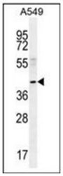
- Experimental details
- Western blot analysis of NEU2 Antibody (N-term) Cat.-No AP52855PU-N in A549 cell line lysates (35ug/lane). This demonstrates the NEU2 antibody detected the NEU2 protein (arrow).

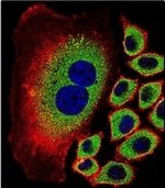
Supportive validation
- Submitted by
- antibodies-online (provider)
- Main image

- Experimental details
- Confocal immunofluorescent analysis of NEU2 Antibody (N-term) Cat.-No AP52855PU-N with A549 cell followed by Alexa Fluor 488-conjugated goat anti-rabbit lgG (green).Actin filaments have been labeled with Alexa Fluor 555 phalloidin (red). DAPI was used to stain the cell nuclear (blue).
Supportive validation
- Submitted by
- antibodies-online (provider)
- Main image
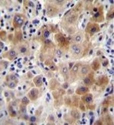
- Experimental details
- Immunohistochemistry analysis in formalin fixed and paraffin embedded human liver tissue reacted with NEU2 Antibody (N-term) Cat.-No AP52855PU-N, which was peroxidase conjugated to the secondary antibody and followed by DAB staining. This data demonstrates the use of NEU2 Antibody (N-term) for immunohistochemistry. Clinical relevance has not been evaluated.

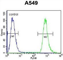
Supportive validation
- Submitted by
- antibodies-online (provider)
- Main image

- Experimental details
- Flow cytometric analysis of A549 cells using NEU2 Antibody (N-term) Cat.-No AP52855PU-N (right histogram) compared to a negative control cell (left histogram). FITC-conjugated goat-anti-rabbit secondary antibodies were used for the analysis.