 Explore
 Validate
 Learn
Learn Western blot
 Immunocytochemistry
Antibody data
- Antibody Data
- Antigen structure
- References [0]
- Comments [0]
- Validations
- Western blot [1]
Submit
Validation data
Reference
Comment
Report error
- Product number
- BS2078 - Provider product page

- Provider
- Bioworld Technology, Inc
- Proper citation
- Bioworld Technology Cat#BS2078, RRID:AB_1663569
- Product name
- DGK Ñ´ (S66) polyclonal antibody
- Antibody type
- Polyclonal
- Antigen
- Synthetic peptide, corresponding to amino acids 34-88 of Human DGK-Ñ´.
- Description
- Diacylglycerol kinases (DGKs) phosphorylate diacylglycerol (DAG) to produce phosphatidic acid. DAG and phosphatidic acid are lipids that act as second messengers in signaling cascades. DGK-Ï« influences cell activation and secretion of lethal exosomes, which in turn control cell death. DGK-ÉÇ is abundant in restricted brain regions such as the caudate putamen and olfactory tubercle. DGK-ÎÛ encodes full-length and truncated transcripts that are present in a range of human tissues, with greatest expression observed in retina. DGK-Ñ´ is most abundant in skeletal muscle. DGK-÷ shows specificity for arachidonylcontaining diacylglycerol and is expressed predominantly in testis. DGK-IJ is most abundant in the cerebellum and hippocampus. DGK-êò is present in brain and retina as a predominant transcript of more than 12 kb, including a long 3-prime untranslated region, with additional low abundance transcripts of 9.5 and 7.5 kb. DGK-»Ò is closely related to DGK-Ñ´. DGK-ø is most abundant in brain and muscle. DGKs have structural motifs that play regulatory roles, and these motifs form the basis for dividing the DGKs into five subtypes.
- Reactivity
- Human, Mouse, Rat
- Host
- Rabbit
- Isotype
- IgG
- Vial size
- 100ul
- Concentration
- 1 mg/ml
- Storage
- Store at 4°C short term. Aliquot and store at -20°C long term. Avoid freeze-thaw cycles.
No comments: Submit comment
Supportive validation
- Submitted by
- Bioworld Technology, Inc (provider)
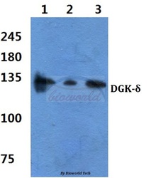
- Main image
- Experimental details
- Western blot (WB) analysis of DGK-¦Ä (S66) pAb at 1:500 dilutionLane1:Hela cell lysateLane2:Raw264.7 cell lysateLane3:PC12 cell lysate
