Explore
 Validate
 Learn
Learn Immunohistochemistry
Antibody data
- Antibody Data
- Antigen structure
- References [3]
- Comments [0]
- Validations
- Immunohistochemistry [2]
- Other assay [3]
Submit
Validation data
Reference
Comment
Report error
- Product number
- PA5-18315 - Provider product page

- Provider
- Invitrogen Antibodies
- Product name
- MRP4 Polyclonal Antibody
- Antibody type
- Polyclonal
- Antigen
- Synthetic peptide
- Description
- This antibody is predicted to react with mouse based on sequence homology. This antibody is tested in Peptide ELISA: antibody detection limit dilution 32,000.
- Reactivity
- Human
- Host
- Goat
- Isotype
- IgG
- Vial size
- 100 μg
- Concentration
- 0.5 mg/mL
- Storage
- -20°C, Avoid Freeze/Thaw Cycles
Submitted references Isothiocyanates (ITCs) 1-(Isothiocyanatomethyl)-4-phenylbenzene and 1-Isothiocyanato-3,5-bis(trifluoromethyl)benzene-Aldehyde Dehydrogenase (ALDH) Inhibitors, Decreases Cisplatin Tolerance and Migratory Ability of NSCLC.
Implications of ABCC4-Mediated cAMP Eflux for CRC Migration.
Knockdown of RAP2A gene expression suppresses cisplatin resistance in gastric cancer cells.
Kryczka J, Kryczka J, Janczewski Ł, Gajda A, Frączyk A, Boncela J, Kolesińska B, Brzeziańska-Lasota E
International journal of molecular sciences 2022 Aug 3;23(15)
International journal of molecular sciences 2022 Aug 3;23(15)
Implications of ABCC4-Mediated cAMP Eflux for CRC Migration.
Kryczka J, Sochacka E, Papiewska-Pająk I, Boncela J
Cancers 2020 Nov 27;12(12)
Cancers 2020 Nov 27;12(12)
Knockdown of RAP2A gene expression suppresses cisplatin resistance in gastric cancer cells.
Zhang J, Wei Y, Min J, Wang Y, Yin L, Cao G, Shen H
Oncology letters 2020 Jan;19(1):350-358
Oncology letters 2020 Jan;19(1):350-358
No comments: Submit comment
Supportive validation
- Submitted by
- Invitrogen Antibodies (provider)
- Main image
- Experimental details
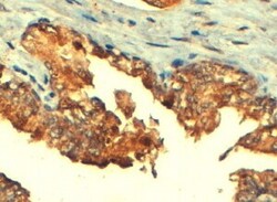
- Immunohistochemistry (PFA fixed) analysis of MRP4 using MRP4 Polyclonal Antibody (Product # PA5-18315) (4 µg/mL) in staining of paraffin embedded Human Prostate. Steamed antigen retrieval with citrate buffer pH 6, HRP-staining.
- Submitted by
- Invitrogen Antibodies (provider)
- Main image
- Experimental details
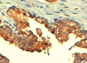
- Immunohistochemistry analysis of MRP4 in human prostate. Samples were incubated with MRP4 polyclonal antibody (Product # PA5-18315) using a dilution of 5 µg/mL. Formalin-fixed, paraffin-embedded tissue after heat-induced antigen retrieval.

Supportive validation
- Submitted by
- Invitrogen Antibodies (provider)
- Main image

- Experimental details
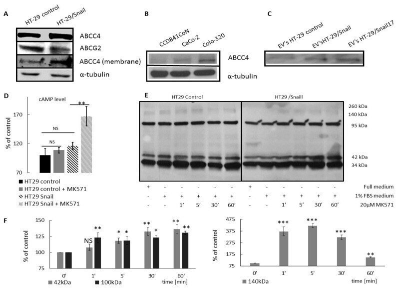
- Figure 3 ABCC4 protein expression level in CRC cell lines. Western blot performed in standard reducing SDS PAGE conditions using goat anti ABCC4 (#PA5 18315, Thermo Scientific) and rabbit anti ABCG2 (#ORB 155559 Biorbyt). ( A ) Protein expression level of ABCC4 and ABCG2 in HT-29 stably overexpressing transcription factor Snail (HT29/Snail) and control HT-29. ABCC4 level in the membrane fraction (obtained by biotynylation using EZ-Link Sulfo -NHS-Biotin Thermo Scientific kit) of HT-29 control cells and HT-29 Snail n = 3. ( B ) ABCC4 protein expression level in CRC cells in different states of EMT: CCD841CoN (most epithelial), CaCo-2 (moderate EMT), and Colo-320 (most mesenchymal) n = 3. ( C ) ABCC4 protein abundance in Extracellular Vesicles (EVs) released from HT-29 control cells and two HT-29 stably overexpressing transcription factor Snail clones (HT-29/Snail and HT-29/Snail17), n = 2. ( D ) Intracellular cAMP level measurement. Accumulation of cAMP in HT29 cells was measured using a cAMP competitive kit (#581001 Cayman Chemical). Cells were incubated for 24 h with MK571 20 uM, or untreated ones were assayed according to the manufacturer's protocol. Calculation were conducted using the Cayman data sheet. cAMP concentration of HT29 was set as 100%. T -test performed, n = 5; * p < 0.05; ** p < 0.005; *** p < 0.001. NS--not statistically significant. ( E ) PKA phosphorylation profile analysis. HT29 Snail cells were seeded on a 6-well plate. Then, 24 h after, full growth mediu
- Submitted by
- Invitrogen Antibodies (provider)
- Main image
- Experimental details
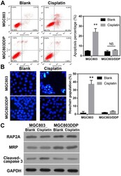
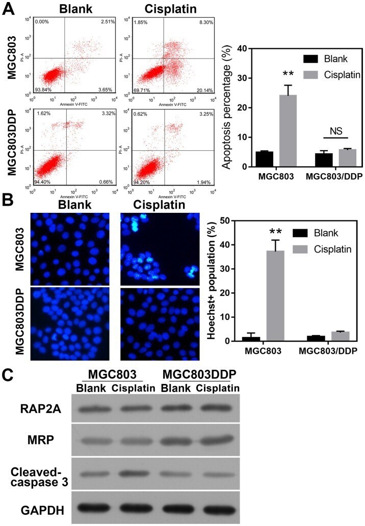
- Figure 2. DDP-resistant gastric cancer cells exhibit suppressed levels of apoptosis and DNA damage. (A) Percentages of apoptotic MGC803 and MGC803/DDP cells following DDP treatment. Cell apoptosis following DDP treatment (1.8 µg/mL for 48 h) was evaluated via flow cytometry. (B) DNA damage in MGC803 and MGC803/DDP cells treated with DDP. DNA damage following DDP treatment (1.8 µg/mL for 48 h) was analyzed by Hoechst 33342 staining and flow cytometry. (C) Levels of RAP2A, MRP and cleaved-caspase-3 in MGC803 and MGC803/DDP cells treated with DDP. Relative levels of protein expression were determined via western blotting, using GAPDH as an internal standard. **P
- Submitted by
- Invitrogen Antibodies (provider)
- Main image
- Experimental details
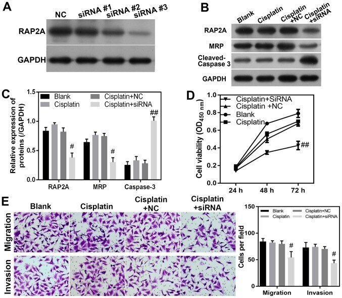
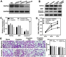
- Figure 3. RAP2A promotes migration, invasion and DDP resistance in MGC803/DDP cells. (A) RAP2A expression was knocked down by siRNA. (B and C) Levels of RAP2A, MRP and cleaved-caspase-3 in RAP2A-knockdown MGC803/DDP cells during DDP treatment. Relative levels of protein expression were determined via western blotting, with GAPDH serving as an internal standard. (D) Effect of RAP2A siRNA knockdown on MGC803/DDP cell viability during DDP treatment. Cell viability was measured using the Cell Counting Kit-8 assay. (E) Migration and invasion of MGC803/DDP cells with RAP2A siRNA knockdown during DDP treatment for 24 h (x100 magnification). Cell migration and invasion were analyzed using the Transwell system. Statistical analysis of MGC803/DDP cell migration and invasion. # P