Explore
 Validate
 Learn
Learn Western blot
 ELISA
ELISAAntibody data
- Antibody Data
- Antigen structure
- References [3]
- Comments [0]
- Validations
- Western blot [2]
- Immunocytochemistry [1]
- Immunohistochemistry [1]
- Other assay [1]
Submit
Validation data
Reference
Comment
Report error
- Product number
- PA1-002 - Provider product page

- Provider
- Invitrogen Antibodies
- Product name
- Huntingtin Polyclonal Antibody
- Antibody type
- Polyclonal
- Antigen
- Synthetic peptide
- Description
- PA1-002 has been successfully used in ELISA, Western Blot, immunofluorescence, and immunohistochemistry applications with human and mouse samples. Neoepitope antibodies distinguish smaller cleaved fragments or processed forms of proteins versus the intact full-length or precursor by using a designed peptide purification process to maximize immunoreactivity to a specific cleavage site. Human HTT caspase cleavage sites generate fragment-specific forms of the protein. Caspase-3/7 has been shown to generate cleavage sites at animo acids 513 and 552. Caspase-2 cleaves at amino acid 552 and caspase-6 at amino acid 586. Neo-specific antibody PA1-002 recognizes the 513 cleaved fragment without detecting the full-length form.
- Reactivity
- Human, Mouse
- Host
- Rabbit
- Isotype
- IgG
- Vial size
- 100 µL
- Concentration
- 0.13 mg/mL
- Storage
- -20°C
Submitted references Identification and evaluation of small molecule pan-caspase inhibitors in Huntington's disease models.
Specific caspase interactions and amplification are involved in selective neuronal vulnerability in Huntington's disease.
Caspase cleavage of mutant huntingtin precedes neurodegeneration in Huntington's disease.
Leyva MJ, Degiacomo F, Kaltenbach LS, Holcomb J, Zhang N, Gafni J, Park H, Lo DC, Salvesen GS, Ellerby LM, Ellman JA
Chemistry & biology 2010 Nov 24;17(11):1189-200
Chemistry & biology 2010 Nov 24;17(11):1189-200
Specific caspase interactions and amplification are involved in selective neuronal vulnerability in Huntington's disease.
Hermel E, Gafni J, Propp SS, Leavitt BR, Wellington CL, Young JE, Hackam AS, Logvinova AV, Peel AL, Chen SF, Hook V, Singaraja R, Krajewski S, Goldsmith PC, Ellerby HM, Hayden MR, Bredesen DE, Ellerby LM
Cell death and differentiation 2004 Apr;11(4):424-38
Cell death and differentiation 2004 Apr;11(4):424-38
Caspase cleavage of mutant huntingtin precedes neurodegeneration in Huntington's disease.
Wellington CL, Ellerby LM, Gutekunst CA, Rogers D, Warby S, Graham RK, Loubser O, van Raamsdonk J, Singaraja R, Yang YZ, Gafni J, Bredesen D, Hersch SM, Leavitt BR, Roy S, Nicholson DW, Hayden MR
The Journal of neuroscience : the official journal of the Society for Neuroscience 2002 Sep 15;22(18):7862-72
The Journal of neuroscience : the official journal of the Society for Neuroscience 2002 Sep 15;22(18):7862-72
No comments: Submit comment
Supportive validation
- Submitted by
- Invitrogen Antibodies (provider)
- Main image
- Experimental details
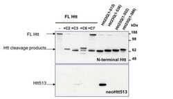
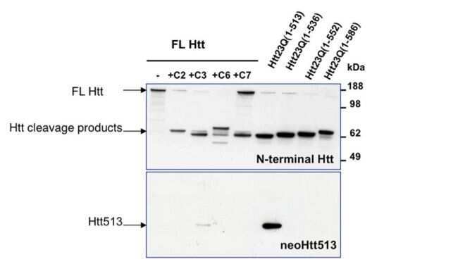
- Western blot analysis of endogenous HTT lysates with or without different caspase activity (Lanes 1-5) and overexpressed recombinant HTT fragment lysates (Lanes 6-9) was performed by loading 20 µg of lysate per well onto a 4-12% Bis-Tris polyacrylamide gel. Proteins were transferred to a nitrocellulose membrane and blocked with 3% BSA/TBST for at least 1 hour. Membranes were then probed with a N-terminal pan-HTT antibody (top panel) or neoepitope-specific rabbit polyclonal antibody Product # PA1-002 (bottom panel) at a dilution of 1:500 overnight at 4°C on a rocking platform. Membranes were then washed in TBS-0.1%Tween 20 and probed with a goat anti-rabbit-HRP secondary antibody at 1:30,000 for at least one hour. Membranes were washed and chemiluminescent detection was performed using SuperSignal West Pico (Product # 34080).
- Submitted by
- Invitrogen Antibodies (provider)
- Main image
- Experimental details
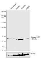
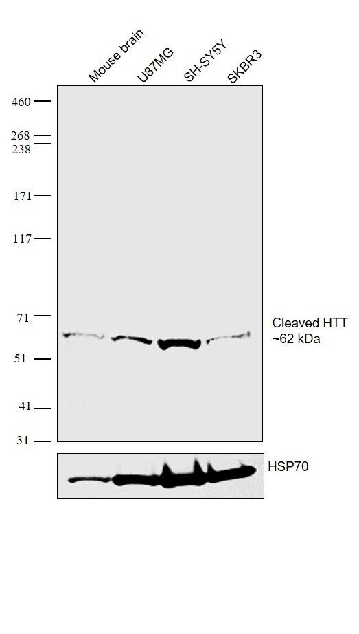
- Western blot was performed using Anti-Huntingtin Polyclonal Antibody (Product # PA1-002) and a 62 kDa band corresponding to cleaved fragment of Huntingtin was observed in Mouse brain tissue lysate and across cell lines tested. Tissue/ whole cell extracts (30 µg lysate) of Mouse Brain (Lane 1), U87MG (Lane 2), SH-SY5Y (Lane 3) and SKBR3 (Lane 4) were electrophoresed using NuPAGE™ 3-8% Tris-Acetate gel (Product # EA0375BOX). Resolved proteins were then transferred onto a nitrocellulose membrane (Product #) by iBlot® 2 Dry Blotting System (Product # IB21001). The bot was probed with the primary antibody (1:1000 dilution) and detected by chemiluminescence with Goat anti-Rabbit IgG (H+L) Superclonal™ Recombinant Secondary Antibody, HRP (Product # A27036, 1:4000 dilution) using the iBright FL 1000 (Product # A32752). Chemiluminescent detection was performed using Novex® ECL Chemiluminescent Substrate Reagent Kit (Product # WP20005).
Supportive validation
- Submitted by
- Invitrogen Antibodies (provider)
- Main image

- Experimental details
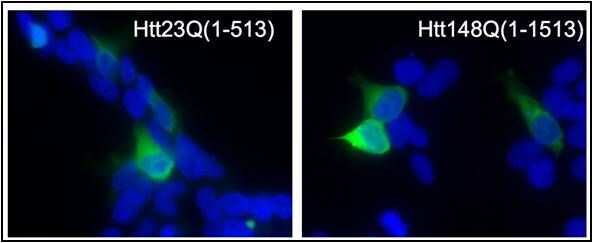
- Immunofluorescent analysis of Caspase cleaved Htt (green) in 293T cells either transfected with an Htt23Q (left panel) or Htt148Q (right panel) stop construct ending in amino acid 513. Formalin fixed cells were permeabilized with 0.1% Triton X-100 in TBS for 10 minutes at room temperature and blocked with 5% normal goat serum for 15 minutes at room temperature. Cells were probed with an Htt neo-epitope 513 polyclonal antibody (Product # PA1-002) at a dilution of 1:50 for at least 1 hour at room temperature, washed with PBS, and incubated with goat-anti-rabbit secondary antibody at room temperature. Nuclei (blue) were stained with Hoechst dye.
Supportive validation
- Submitted by
- Invitrogen Antibodies (provider)
- Main image
- Experimental details
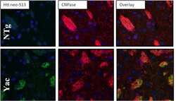
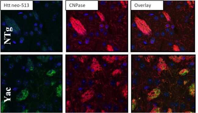
- Immunohistochemistry was performed on tissues from either transgenic HD mice of the YAC128 line (bottom panels) or their WT littermates (top panels). Striatum tissue samples were probed with an Htt neo-epitope 513 polyclonal antibody (Product # PA1-002) at a dilution of 1:50 overnight at 4°C in a humidified chamber. Tissues were washed extensively and endogenous peroxidase activity quenched for 30 minutes at room temperature. Detection was performed using a goat anti-rabbit HRP secondary antibody (green). Tissues were counterstained with CNPase (red) and nuclei stain (blue) and prepped for mounting.
Supportive validation
- Submitted by
- Invitrogen Antibodies (provider)
- Main image
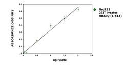
- Experimental details
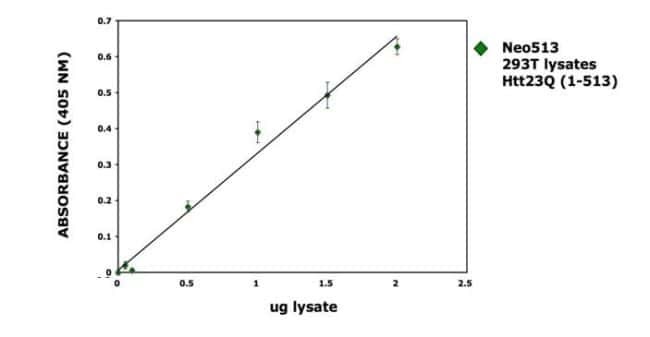
- Sandwich ELISA was performed with a monoclonal antibody to Htt and an Htt neo-epitope 513 polyclonal antibody (Product # PA1-002) to determine the antigen concentration of the Htt cleavage products. The curve represents a dose response for neo513 in 293T cells overexpressing the Htt construct.