Explore
 Validate
 Learn
LearnPA5-30507
antibody from Invitrogen Antibodies
Targeting: TRAF3IP1
DKFZP434F124, FAP116, IFT54, MIP-T3, MIPT3
 Western blot
Antibody data
- Antibody Data
- Antigen structure
- References [2]
- Comments [0]
- Validations
- Western blot [3]
- Immunohistochemistry [1]
- Other assay [2]
Submit
Validation data
Reference
Comment
Report error
- Product number
- PA5-30507 - Provider product page

- Provider
- Invitrogen Antibodies
- Product name
- TRAF3IP1 Polyclonal Antibody
- Antibody type
- Polyclonal
- Antigen
- Recombinant full-length protein
- Description
- Recommended positive controls: U87-MG, mouse testis, rat testis. Predicted reactivity: Bovine (85%). Store product as a concentrated solution. Centrifuge briefly prior to opening the vial.
- Reactivity
- Human, Mouse, Rat
- Host
- Rabbit
- Isotype
- IgG
- Vial size
- 100 μL
- Concentration
- 1 mg/mL
- Storage
- Store at 4°C short term. For long term storage, store at -20°C, avoiding freeze/thaw cycles.
Submitted references Mutations in GRK2 cause Jeune syndrome by impairing Hedgehog and canonical Wnt signaling.
Mutations in DYNC2LI1 disrupt cilia function and cause short rib polydactyly syndrome.
Bosakova M, Abraham SP, Nita A, Hruba E, Buchtova M, Taylor SP, Duran I, Martin J, Svozilova K, Barta T, Varecha M, Balek L, Kohoutek J, Radaszkiewicz T, Pusapati GV, Bryja V, Rush ET, Thiffault I, Nickerson DA, Bamshad MJ, University of Washington Center for Mendelian Genomics, Rohatgi R, Cohn DH, Krakow D, Krejci P
EMBO molecular medicine 2020 Nov 6;12(11):e11739
EMBO molecular medicine 2020 Nov 6;12(11):e11739
Mutations in DYNC2LI1 disrupt cilia function and cause short rib polydactyly syndrome.
Taylor SP, Dantas TJ, Duran I, Wu S, Lachman RS, University of Washington Center for Mendelian Genomics Consortium, Nelson SF, Cohn DH, Vallee RB, Krakow D
Nature communications 2015 Jun 16;6:7092
Nature communications 2015 Jun 16;6:7092
No comments: Submit comment
Supportive validation
- Submitted by
- Invitrogen Antibodies (provider)
- Main image
- Experimental details
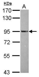
- Western Blot using TRAF3IP1 Polyclonal Antibody (Product # PA5-30507). Sample (30 µg of whole cell lysate). Lane A: U87-MG. 7.5% SDS PAGE. TRAF3IP1 Polyclonal Antibody (Product # PA5-30507) diluted at 1:3,000.

- Submitted by
- Invitrogen Antibodies (provider)
- Main image
- Experimental details
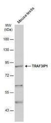
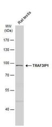
- Western Blot analysis of TRAF3IP1 was performed by separating 50 µg of Mouse tissue extracts by 7.5% SDS-PAGE. Proteins were transferred to a membrane and probed with a TRAF3IP1 Polyclonal Antibody (Product # PA5-30507) at a dilution of 1:500. The HRP-conjugated anti-rabbit IgG antibody was used to detect the primary antibody.
- Submitted by
- Invitrogen Antibodies (provider)
- Main image
- Experimental details
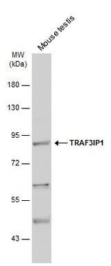
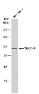
- Western Blot analysis of TRAF3IP1 was performed by separating 50 µg of Rat tissue extracts by 7.5% SDS-PAGE. Proteins were transferred to a membrane and probed with a TRAF3IP1 Polyclonal Antibody (Product # PA5-30507) at a dilution of 1:500. The HRP-conjugated anti-rabbit IgG antibody was used to detect the primary antibody.
Supportive validation
- Submitted by
- Invitrogen Antibodies (provider)
- Main image
- Experimental details
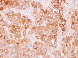
- Immunohistochemical analysis of paraffin-embedded H441 xenograft, using TRAF3IP1 (Product # PA5-30507) antibody at 1:100 dilution. Antigen Retrieval: Citrate buffer, pH 6.0, 15 min.

Supportive validation
- Submitted by
- Invitrogen Antibodies (provider)
- Main image
- Experimental details
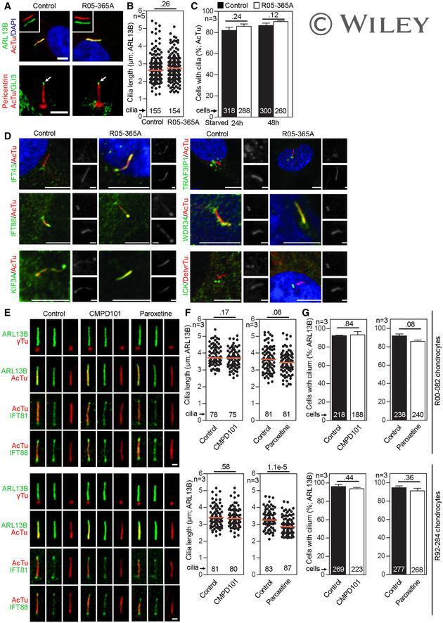
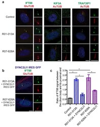
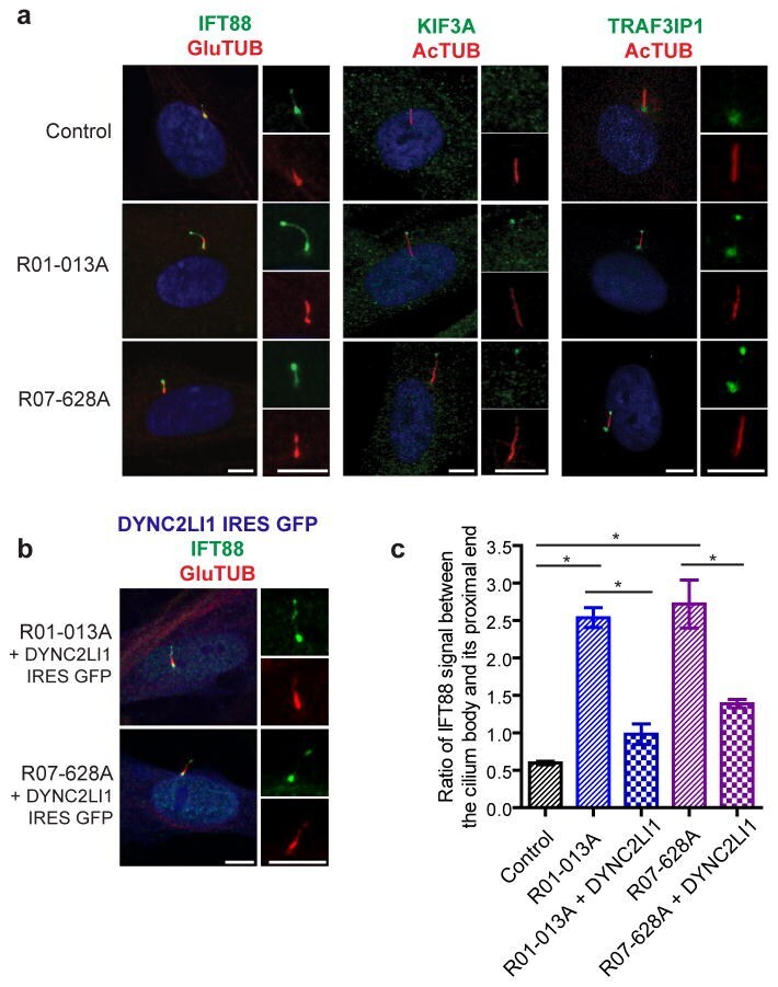
- Figure 6 DYNC2LI1 mutations delay retrograde IFT and lead to the ciliary accumulation of IFT components. (a) Immunofluorescence micrographs of control and SRPS cells stained as labeled. Cilia (2x) are shown on the right. (b) Expression of untagged wild type DYNC2LI1 through an IRES-GFP vector (artificially colored blue) rescues ciliary accumulation of IFT components. (c) Quantification of IFT88 signal: graph shows the mean ratio of IFT88 signal between the cilium body and its proximal end +- s.e.m (n=20 X 3 independent experiments). Statistical analyses performed using Mann-Whitney test, p
- Submitted by
- Invitrogen Antibodies (provider)
- Main image
- Experimental details
- EV1 Figure GRK 2 is not required for ciliogenesis or IFT A-C Primary cilia are normal in GRK2 -/- fibroblasts. Cells were serum starved for 24 or 48 h to induce cilia, and (A) immunostained for acetylated tubulin (AcTu, red), ARL13B (green) (upper panel; insets show the individual signals), or AcTu (red), pericentrin (red) and GLI3 (green) (lower panel). Control and GRK2 -/- cilia were positive for AcTu and ARL13B, and showed similar localization of GLI3 to the tips (lower panel, arrows). Acetylated tubulin and pericentrin staining were used to visualize the axoneme and centrioles, respectively. Scale bar, 2 mum. (B) Length distribution of cilia did not differ between control and GRK2 -/- cells. Red bars show medians; dots represent individual cilia. Mann-Whitney U test; number of biological experiments and the total numbers of analyzed cilia are indicated. (C) There was no difference in the efficiency of ciliogenesis between control and GRK2 -/- cells. Mean +- SEM. Welch's t -test; number of biological experiments and the total numbers of analyzed cells are indicated. D Loss of GRK2 did not affect localization of ciliary and IFT components. Immunofluorescence of serum-starved (48 hr) control and GRK2 -/- fibroblasts immunostained with AcTu or detyrosinated tubulin (DetyrTu) to mark the cilium (red), and IFT43, IFT88, KIF3A, TRAF3IP1, WDR34, or ICK (green). No significant differences in staining were found between control and GRK2 -/- cilia, suggesting normal IFT. Scale bars,