Explore
 Validate
 Learn
Learn Immunocytochemistry
 Immunohistochemistry
Antibody data
- Antibody Data
- Antigen structure
- References [1]
- Comments [0]
- Validations
- Immunocytochemistry [2]
- Flow cytometry [3]
Submit
Validation data
Reference
Comment
Report error
- Product number
- GLUT1-G100 - Provider product page

- Provider
- METAFORA biosystems
- Product name
- Anti-GLUT1 RBD
- Antibody type
- Recombinant
- Antigen
- Other
- Description
- GFP fusion recombinant protein purified from CHO supernatant
- Reactivity
- Human, Mouse, Rat
- Antigen sequence
QYVEQLC- Epitope
- Binds to an epitope located within extracellular loop 6 of Glut1 as determined by directed mutagenesis
- Vial size
- 300 µl
- Concentration
- 0.1 mg/ml
- Storage
- store in -20°C freezer. Working dilution samples should be discarded if not used within 12 hours.
- Handling
- The antibody solution should be gently mixed before use.
Submitted references HIV-1 pathogenicity and virion production are dependent on the metabolic phenotype of activated CD4+ T cells.
Hegedus A, Kavanagh Williamson M, Huthoff H
Retrovirology 2014 Nov 25;11(1):98
Retrovirology 2014 Nov 25;11(1):98
No comments: Submit comment
Supportive validation
Supportive validation
- Submitted by
- METAFORA biosystems (provider)
- Main image
- Experimental details
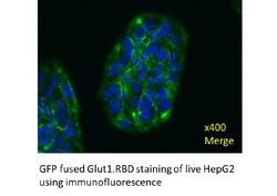
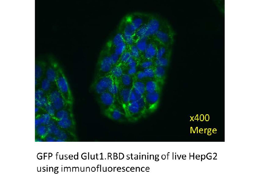
- Immunofluorescent staining of human cell line HepG2 shows cell surface staining
- Sample type
- HepG2
- Validation comment
- The subcellular location is supported by literature.
- Protocol
- Protocol
Supportive validation
- Submitted by
- METAFORA biosystems (provider)
- Main image

- Experimental details
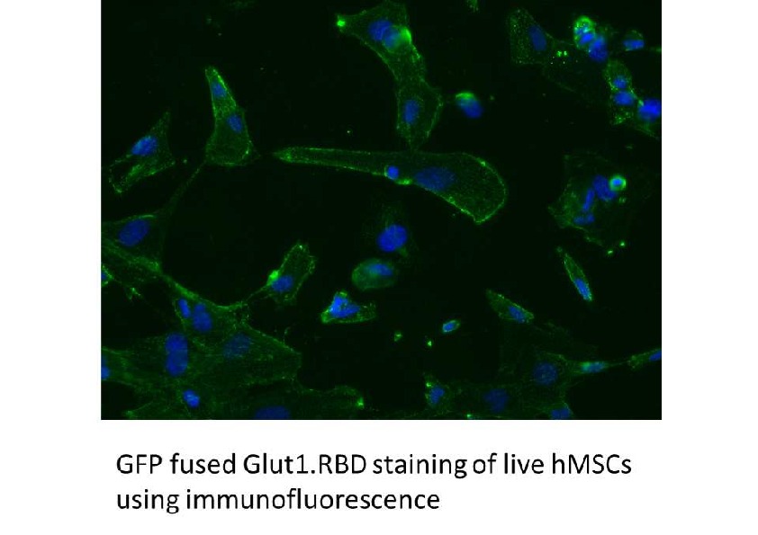
- Immunofluorescent staining of human Mesenchymal Stem Cells shows cell surface staining
- Sample type
- hMSC
- Validation comment
- The subcellular location is supported by literature.
- Protocol
- Protocol
Supportive validation
Supportive validation
Supportive validation
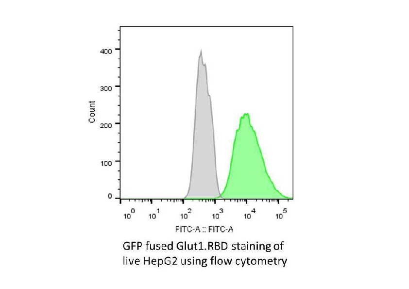
- Submitted by
- METAFORA biosystems (provider)
- Main image

- Experimental details
- FACS fluorescent shift observed for live non permeabilized human cell line HepG2
- Sample type
- HepG2
- Validation comment
- The subcellular location is supported by literature.
- Protocol
- Protocol
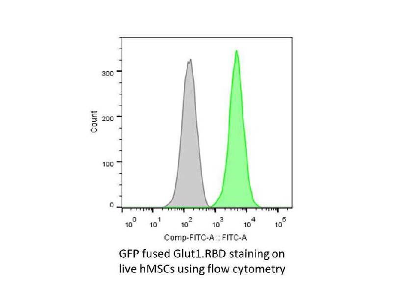
Supportive validation
- Submitted by
- METAFORA biosystems (provider)
- Main image
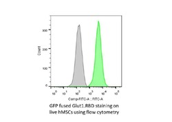
- Experimental details
- FACS fluorescent shift observed for live non permeabilized human Mesenchymal Stem Cells
- Sample type
- hMSCs
- Validation comment
- The subcellular location is supported by literature.
- Protocol
- Protocol
Supportive validation
- Submitted by
- METAFORA biosystems (provider)
- Main image
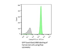
- Experimental details
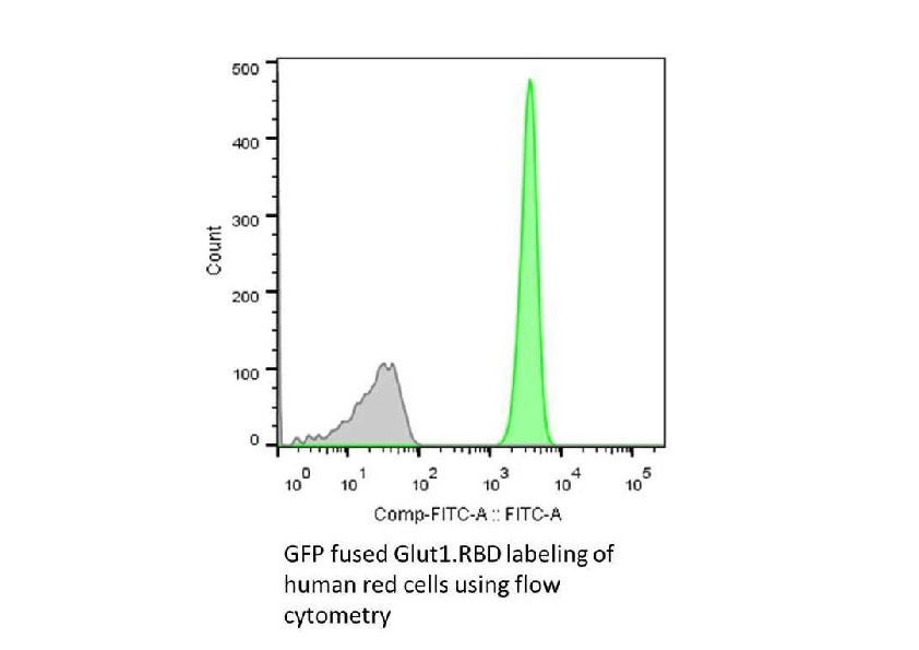
- FACS fluorescent shift observed for live non permeabilized human Red Cells
- Sample type
- h Red Cells
- Validation comment
- The subcellular location is supported by literature.
- Protocol
- Protocol