Explore
 Validate
 Learn
Learn Immunocytochemistry
Antibody data
- Antibody Data
- Antigen structure
- References [5]
- Comments [0]
- Validations
- Immunocytochemistry [1]
- Immunohistochemistry [1]
Submit
Validation data
Reference
Comment
Report error
- Product number
- HPA031345 - Provider product page

- Provider
- Atlas Antibodies
- Proper citation
- Atlas Antibodies Cat#HPA031345, RRID:AB_2673835
- Product name
- Anti-SLC2A1
- Antibody type
- Polyclonal
- Description
- Polyclonal Antibody against Human SLC2A1, Gene description: solute carrier family 2 (facilitated glucose transporter), member 1, Alternative Gene Names: DYT18, GLUT, GLUT1, HTLVR, Validated applications: IHC, ICC, Uniprot ID: P11166, Storage: Store at +4°C for short term storage. Long time storage is recommended at -20°C.
- Reactivity
- Human, Mouse
- Host
- Rabbit
- Conjugate
- Unconjugated
- Isotype
- IgG
- Vial size
- 100 µl
- Concentration
- 0.3 mg/ml
- Storage
- Store at +4°C for short term storage. Long time storage is recommended at -20°C.
- Handling
- The antibody solution should be gently mixed before use.
Submitted references Endothelial DR6 in blood-brain barrier malfunction in Alzheimer’s disease
An easy-to-perform method for microvessel isolation and primary brain endothelial cell culture to study Alzheimer's disease
Activation of Wnt/β-catenin pathway mitigates blood–brain barrier dysfunction in Alzheimer’s disease
Brain capillary pericytes are metabolic sentinels that control blood flow through a K(ATP) channel-dependent energy switch.
Transcriptome Response and Spatial Pattern of Gene Expression in the Primate Subventricular Zone Neurogenic Niche After Cerebral Ischemia
Huang X, Qi J, Su Y, Zhou Y, Wang Q, Huang T, Xue D, Zeng Y, Verkhratsky A, Zhou B, Chen H, Yi C
Cell Death & Disease 2024;15(4)
Cell Death & Disease 2024;15(4)
An easy-to-perform method for microvessel isolation and primary brain endothelial cell culture to study Alzheimer's disease
Chen Y, Huang X, Chen H, Yi C
Heliyon 2024;10(12):e33077
Heliyon 2024;10(12):e33077
Activation of Wnt/β-catenin pathway mitigates blood–brain barrier dysfunction in Alzheimer’s disease
Wang Q, Huang X, Su Y, Yin G, Wang S, Yu B, Li H, Qi J, Chen H, Zeng W, Zhang K, Verkhratsky A, Niu J, Yi C
Brain 2022;145(12):4474-4488
Brain 2022;145(12):4474-4488
Brain capillary pericytes are metabolic sentinels that control blood flow through a K(ATP) channel-dependent energy switch.
Hariharan A, Robertson CD, Garcia DCG, Longden TA
Cell reports 2022 Dec 27;41(13):111872
Cell reports 2022 Dec 27;41(13):111872
Transcriptome Response and Spatial Pattern of Gene Expression in the Primate Subventricular Zone Neurogenic Niche After Cerebral Ischemia
Chongtham M, Wang H, Thaller C, Hsiao N, Vachkov I, Pavlov S, Williamson L, Yamashima T, Stoykova A, Yan J, Eichele G, Tonchev A
Frontiers in Cell and Developmental Biology 2020;8
Frontiers in Cell and Developmental Biology 2020;8
No comments: Submit comment
Supportive validation
- Submitted by
- Atlas Antibodies (provider)
- Main image
- Experimental details
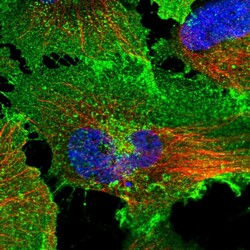
- Immunofluorescent staining of human cell line U-251 MG shows localization to plasma membrane.
- Sample type
- Human

Supportive validation
- Submitted by
- Atlas Antibodies (provider)
- Enhanced method
- Orthogonal validation
- Main image
- Experimental details
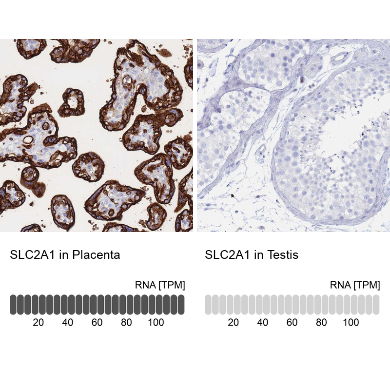
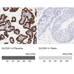
- Immunohistochemistry analysis in human placenta and testis tissues using HPA031345 antibody. Corresponding SLC2A1 RNA-seq data are presented for the same tissues.
- Sample type
- Human
- Protocol
- Protocol