Explore
 Validate
 Learn
LearnMA5-31960
antibody from Invitrogen Antibodies
Targeting: SLC2A1
CSE, DYT18, DYT9, GLUT, GLUT1, HTLVR
 Western blot
Antibody data
- Antibody Data
- Antigen structure
- References [2]
- Comments [0]
- Validations
- Western blot [3]
- Immunocytochemistry [8]
- Immunohistochemistry [7]
- Flow cytometry [2]
- Other assay [1]
Submit
Validation data
Reference
Comment
Report error
- Product number
- MA5-31960 - Provider product page

- Provider
- Invitrogen Antibodies
- Product name
- GLUT1 Recombinant Rabbit Monoclonal Antibody (SA0377)
- Antibody type
- Monoclonal
- Antigen
- Synthetic peptide
- Description
- Recombinant rabbit monoclonal antibodies are produced using in vitro expression systems. The expression systems are developed by cloning in the specific antibody DNA sequences from immunoreactive rabbits. Then, individual clones are screened to select the best candidates for production. The advantages of using recombinant rabbit monoclonal antibodies include: better specificity and sensitivity, lot-to-lot consistency, animal origin-free formulations, and broader immunoreactivity to diverse targets due to larger rabbit immune repertoire.
- Reactivity
- Human, Mouse, Rat
- Host
- Rabbit
- Isotype
- IgG
- Antibody clone number
- SA0377
- Vial size
- 100 μL
- Concentration
- 1 mg/mL
- Storage
- Store at 4°C short term. For long term storage, store at -20°C, avoiding freeze/thaw cycles.
Submitted references The proteomic analysis of breast cell line exosomes reveals disease patterns and potential biomarkers.
Metabolic Deregulation of the Blood-Outer Retinal Barrier in Retinitis Pigmentosa.
Risha Y, Minic Z, Ghobadloo SM, Berezovski MV
Scientific reports 2020 Aug 11;10(1):13572
Scientific reports 2020 Aug 11;10(1):13572
Metabolic Deregulation of the Blood-Outer Retinal Barrier in Retinitis Pigmentosa.
Wang W, Kini A, Wang Y, Liu T, Chen Y, Vukmanic E, Emery D, Liu Y, Lu X, Jin L, Lee SJ, Scott P, Liu X, Dean K, Lu Q, Fortuny E, James R, Kaplan HJ, Du J, Dean DC
Cell reports 2019 Jul 30;28(5):1323-1334.e4
Cell reports 2019 Jul 30;28(5):1323-1334.e4
No comments: Submit comment
Supportive validation
- Submitted by
- Invitrogen Antibodies (provider)
- Main image
- Experimental details
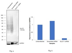
- Knockdown of GLUT1 was achieved by transfecting MCF7 with GLUT1 specific siRNAs (Silencer® select Product # s12927, s12926). Western blot analysis (Fig. a) was performed using Membrane enriched extracts from the GLUT1 knockdown cells (lane 3), non-targeting scrambled siRNA transfected cells (lane 2) and untransfected cells (lane 1). The blot was probed with GLUT1 Recombinant Rabbit Monoclonal Antibody (SA0377) (Product # MA5-31960, 1:1000 ) and Goat anti-Rabbit IgG (Heavy Chain) Superclonal™ Recombinant Secondary Antibody, HRP (Product # A27036, 1:4000). Densitometric analysis of this western blot is shown in histogram (Fig. b). Decrease in signal upon siRNA mediated knock down confirms that antibody is specific to GLUT1. The Target GLUT-1 shows pick up in positive cell models as a streak like pattern. The siRNA lane (Lane 3) shows almost 90% loss of this streak like signal.

- Submitted by
- Invitrogen Antibodies (provider)
- Main image
- Experimental details
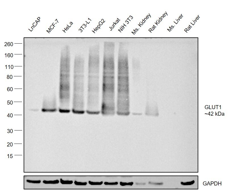
- Western blot was performed using Anti-GLUT1 Recombinant Rabbit Monoclonal Antibody (SA0377) (Product # MA5-31960) and a ~42 kDa band corresponding to GLUT1 was observed across cell lines and tissues tested . Membrane enriched extracts (50 µg lysate) of LNCaP (Lane 1), MCF7 (Lane 2), HeLa (Lane 3), 3T3-L1 (Lane 4), HepG2 (Lane 5), Jurkat (Lane 6), NIH 3T3 (Lane 7), Mouse Kidney (Lane 8), Rat Kidney (Lane 9), Mouse Liver (Lane 10) and Rat Liver (Lane 11) were electrophoresed using NuPAGE™ 4-12% Bis-Tris Protein Gel (Product # NP0321BOX). Resolved proteins were then transferred onto a Nitrocellulose membrane (Product # LC2001) by iBlot® 2 Dry Blotting System (Product # IB21001). The blot was probed with the primary antibody (1:1000 dilution) and detected by chemiluminescence with Goat anti-Rabbit IgG (Heavy Chain) Superclonal™ Recombinant Secondary Antibody, HRP (Product # A27036, 1:4000) using the iBright FL 1000 (Product # A32752). Chemiluminescent detection was performed using Novex® ECL Chemiluminescent Substrate Reagent Kit (Product # WP20005).A streak like pattern was observed in the positive cell lines and tissues which is expected of GLUT-1 target.
- Submitted by
- Invitrogen Antibodies (provider)
- Main image

- Experimental details
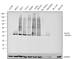
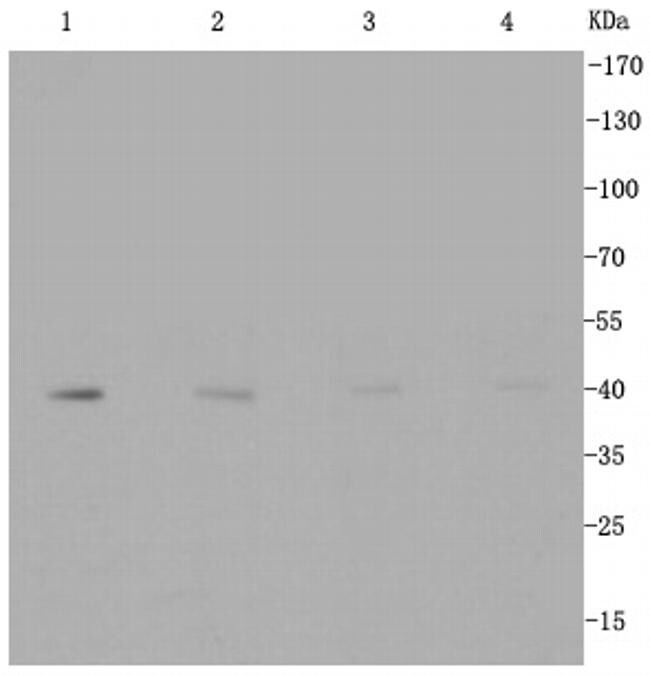
- Western blot analysis of GLUT1 in different lysates using a Monoclonal antibody (Product #MA5-31960) at a dilution of 1:1000. Positive control: Lane 1: Hela, Lane 2: MCF-7, Lane 3: Jurkat, Lane 4: NIH/3T3.
Supportive validation
- Submitted by
- Invitrogen Antibodies (provider)
- Main image
- Experimental details
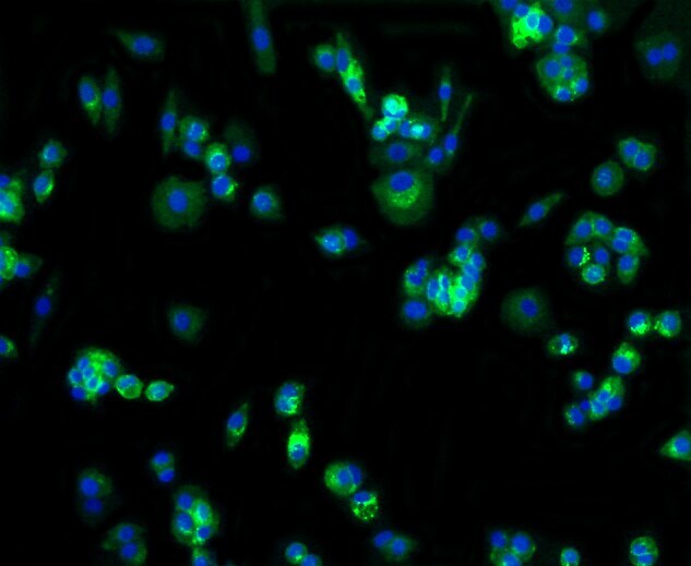
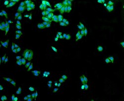
- Immunocytochemical analysis of GLUT1 in Hela cells using a GLUT1 Monoclonal antibody (Product # MA5-31960) as seen in green. The nuclear counter stain is DAPI (blue). Cells were fixed in paraformaldehyde, permeabilised with 0.25% Triton X100/PBS.
- Submitted by
- Invitrogen Antibodies (provider)
- Main image

- Experimental details
- Immunocytochemical analysis of GLUT1 in MCF-7 cells using a GLUT1 Monoclonal antibody (Product # MA5-31960) as seen in green. The nuclear counter stain is DAPI (blue). Cells were fixed in paraformaldehyde, permeabilised with 0.25% Triton X100/PBS.
- Submitted by
- Invitrogen Antibodies (provider)
- Main image
- Experimental details
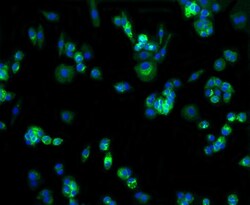
- Immunocytochemical analysis of GLUT1 in HepG2 cells using a GLUT1 Monoclonal antibody (Product # MA5-31960) as seen in green. Cells were fixed in paraformaldehyde, permeabilised with 0.25% Triton X100/PBS.

- Submitted by
- Invitrogen Antibodies (provider)
- Main image
- Experimental details
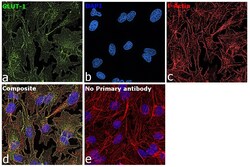
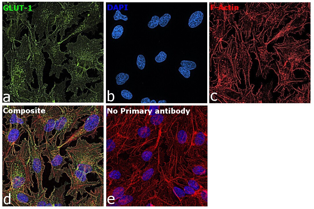
- Immunofluorescence analysis of GLUT1 was performed using 70% confluent log phase Hep G2 cells. The cells were fixed with 4% paraformaldehyde for 10 minutes, permeabilized with 0.1% Triton™ X-100 for 15 minutes, and blocked with 2% BSA for 45 minutes at room temperature. The cells were labeled with GLUT1 Recombinant Rabbit Monoclonal Antibody (SA0377) (Product # MA5-31960) at 1:100 dilution in 0.1% BSA, incubated at 4 degree celsius overnight and then labeled with Donkey anti-Rabbit IgG (H+L) Highly Cross-Adsorbed Secondary Antibody, Alexa Fluor Plus 488 (Product # A32790), 1:2000 dilution, for 45 minutes at room temperature (Panel a: Blue). Nuclei (Panel b:Green) were stained with ProLong™ Diamond Antifade Mountant with DAPI (Product # P36962). F-actin (Panel c: Red) was stained with Rhodamine Phalloidin (Product # R415, 1:300). Panel d represents the merged image showing Plasma membrane and cytoplasm localization. Panel e represents control cells with no primary antibody to assess background. The images were captured at 60X magnification.
- Submitted by
- Invitrogen Antibodies (provider)
- Main image

- Experimental details
- Immunofluorescence analysis of GLUT1 was performed using 70% confluent log phase Hep G2 cells. The cells were fixed with 4% paraformaldehyde for 10 minutes, permeabilized with 0.1% Triton™ X-100 for 15 minutes, and blocked with 2% BSA for 45 minutes at room temperature. The cells were labeled with GLUT1 Recombinant Rabbit Monoclonal Antibody (SA0377) (Product # MA5-31960) at 1:100 dilution in 0.1% BSA, incubated at 4 degree celsius overnight and then labeled with Donkey anti-Rabbit IgG (H+L) Highly Cross-Adsorbed Secondary Antibody, Alexa Fluor Plus 488 (Product # A32790), 1:2000 dilution, for 45 minutes at room temperature (Panel a: Blue). Nuclei (Panel b:Green) were stained with ProLong™ Diamond Antifade Mountant with DAPI (Product # P36962). F-actin (Panel c: Red) was stained with Rhodamine Phalloidin (Product # R415, 1:300). Panel d represents the merged image showing Plasma membrane and cytoplasm localization. Panel e represents control cells with no primary antibody to assess background. The images were captured at 60X magnification.

- Submitted by
- Invitrogen Antibodies (provider)
- Main image

- Experimental details
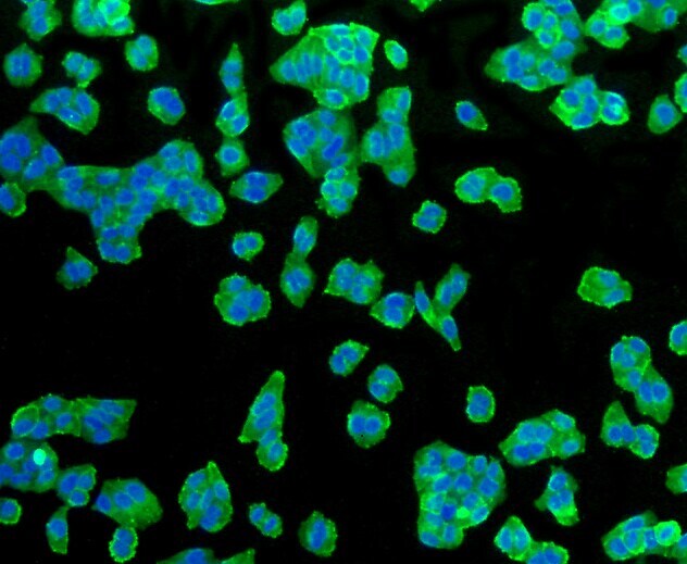
- Immunocytochemical analysis of GLUT1 in HepG2 cells using a GLUT1 Monoclonal antibody (Product # MA5-31960) as seen in green. Cells were fixed in paraformaldehyde, permeabilised with 0.25% Triton X100/PBS.

- Submitted by
- Invitrogen Antibodies (provider)
- Main image
- Experimental details
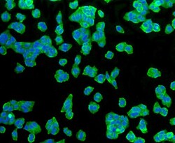
- Immunocytochemical analysis of GLUT1 in MCF-7 cells using a GLUT1 Monoclonal antibody (Product # MA5-31960) as seen in green. The nuclear counter stain is DAPI (blue). Cells were fixed in paraformaldehyde, permeabilised with 0.25% Triton X100/PBS.

- Submitted by
- Invitrogen Antibodies (provider)
- Main image

- Experimental details
- Immunocytochemical analysis of GLUT1 in Hela cells using a GLUT1 Monoclonal antibody (Product # MA5-31960) as seen in green. The nuclear counter stain is DAPI (blue). Cells were fixed in paraformaldehyde, permeabilised with 0.25% Triton X100/PBS.

Supportive validation
- Submitted by
- Invitrogen Antibodies (provider)
- Main image

- Experimental details
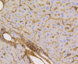
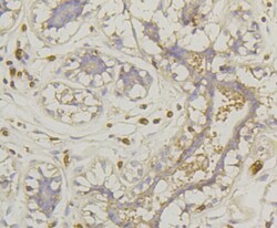
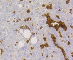
- Immunohistochemical analysis of GLUT1 of paraffin-embedded Human liver tissue using a GLUT1 Monoclonal antibody (Product #MA5-31960). Counter stained with hematoxylin.

- Submitted by
- Invitrogen Antibodies (provider)
- Main image
- Experimental details
- Immunohistochemical analysis of GLUT1 of paraffin-embedded Mouse liver tissue using a GLUT1 Monoclonal antibody (Product #MA5-31960). Counter stained with hematoxylin.

- Submitted by
- Invitrogen Antibodies (provider)
- Main image

- Experimental details
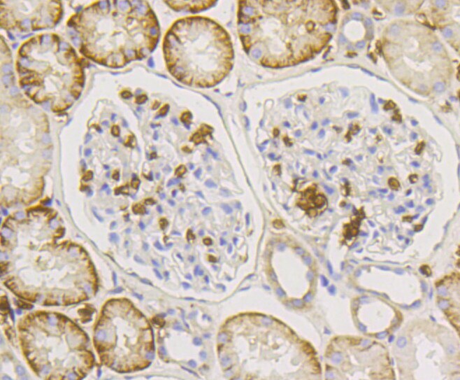
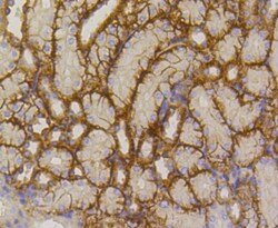
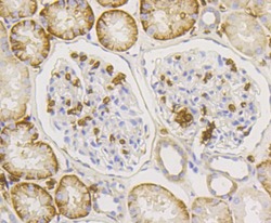
- Immunohistochemical analysis of GLUT1 of paraffin-embedded Human kidney tissue using a GLUT1 Monoclonal antibody (Product #MA5-31960). Counter stained with hematoxylin.
- Submitted by
- Invitrogen Antibodies (provider)
- Main image
- Experimental details
- Immunohistochemical analysis of GLUT1 of paraffin-embedded Human breast carcinoma tissue using a GLUT1 Monoclonal antibody (Product #MA5-31960). Counter stained with hematoxylin.

- Submitted by
- Invitrogen Antibodies (provider)
- Main image
- Experimental details
- Immunohistochemical analysis of GLUT1 of paraffin-embedded Mouse kidney tissue using a GLUT1 Monoclonal antibody (Product #MA5-31960). Counter stained with hematoxylin.

- Submitted by
- Invitrogen Antibodies (provider)
- Main image
- Experimental details
- Immunohistochemical analysis of GLUT1 of paraffin-embedded Human kidney tissue using a GLUT1 Monoclonal antibody (Product #MA5-31960). Counter stained with hematoxylin.

- Submitted by
- Invitrogen Antibodies (provider)
- Main image
- Experimental details
- Immunohistochemical analysis of GLUT1 of paraffin-embedded Human liver tissue using a GLUT1 Monoclonal antibody (Product #MA5-31960). Counter stained with hematoxylin.

Supportive validation
- Submitted by
- Invitrogen Antibodies (provider)
- Main image
- Experimental details
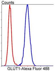
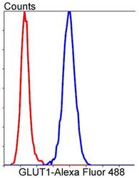
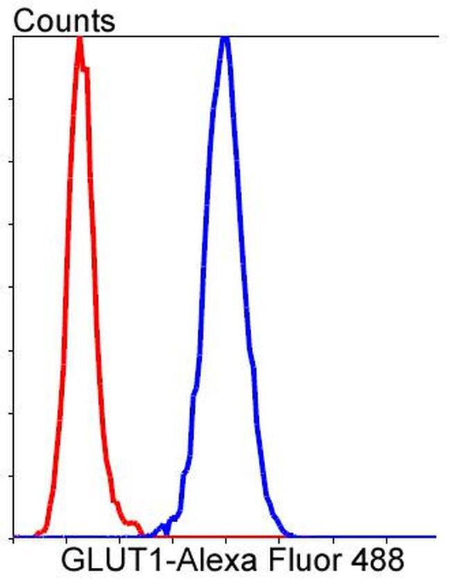
- Flow Cytometric analysis of GLUT1 in Hela cells using a GLUT1 Monoclonal Antibody (Product # MA5-31960) at a dilution of 1:50, as seen in blue compared with an unlabelled control (cells without incubation with primary antibody; red). Alexa Fluor 488-conjugated goat anti rabbit IgG was used as the secondary antibody.
- Submitted by
- Invitrogen Antibodies (provider)
- Main image
- Experimental details
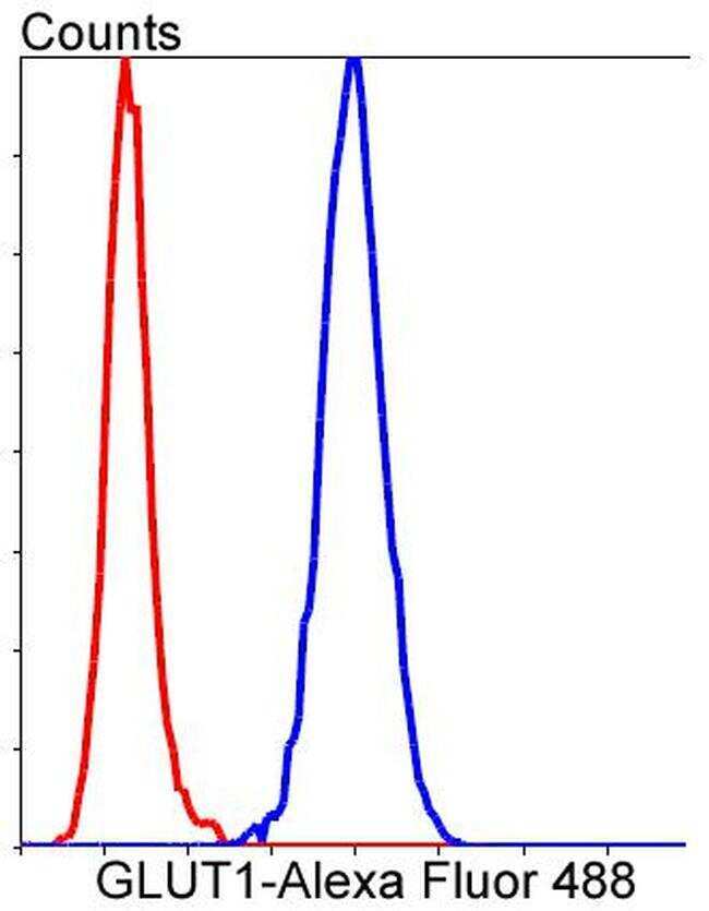
- Flow Cytometric analysis of GLUT1 in Hela cells using a GLUT1 Monoclonal Antibody (Product # MA5-31960) at a dilution of 1:50, as seen in blue compared with an unlabelled control (cells without incubation with primary antibody; red). Alexa Fluor 488-conjugated goat anti rabbit IgG was used as the secondary antibody.
Supportive validation
- Submitted by
- Invitrogen Antibodies (provider)
- Main image
- Experimental details
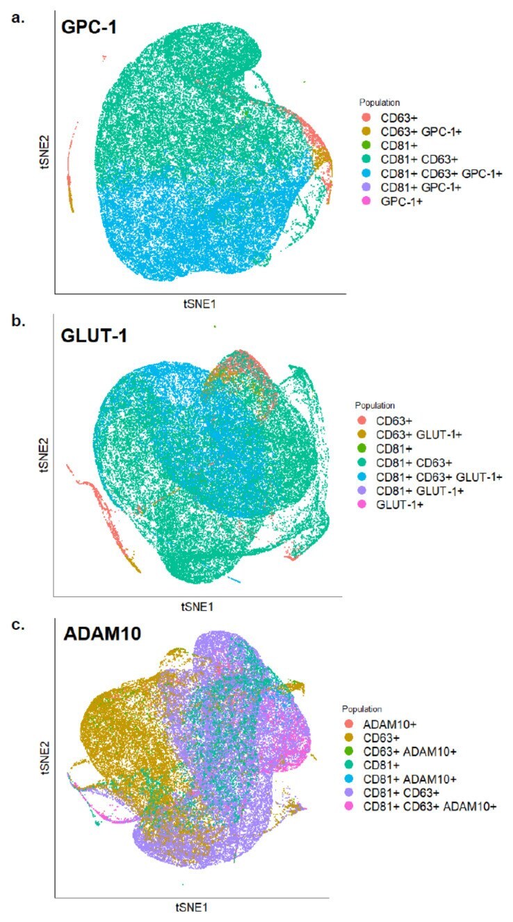
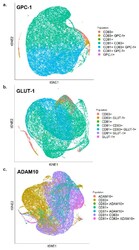
- Figure 5 tSNE plots of exosomes analyzed by FACS showing expression levels of ( a ) GPC-1 ( b ) GLUT-1 and ( c ) ADAM10 along with CD81 and CD63 on the surface of BC-derived exosomes.