Explore
 Validate
 Learn
LearnM00163-1
antibody from Boster Biological Technology
Targeting: SLC2A1
CSE, DYT18, DYT9, GLUT, GLUT1, HTLVR
 Western blot
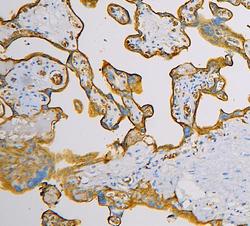
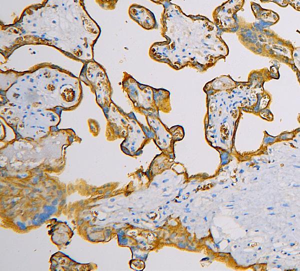
 Immunocytochemistry
Antibody data
- Antibody Data
- Antigen structure
- References [1]
- Comments [0]
- Validations
- Western blot [1]
Submit
Validation data
Reference
Comment
Report error
- Product number
- M00163-1 - Provider product page

- Provider
- Boster Biological Technology
- Product name
- Anti-SLC2A1 Antibody Picoband™ (monoclonal, 10C10)
- Antibody type
- Monoclonal
- Description
- Mouse IgG monoclonal antibody for SLC2A1 detection. Tested with WB, IHC-P, ICC/IF, FCM in Human.
- Reactivity
- Human
- Host
- Mouse
- Isotype
- IgG
- Antibody clone number
- 10C10
- Vial size
- 100μg/vial
- Concentration
- 0.5-1mg/ml, actual concentration vary by lot. Use suggested dilution ratio to decide dilution procedure.
- Storage
- At -20°C for one year. After reconstitution, at 4°C for one month. It can also be aliquoted and stored frozen at -20°C for a longer time. Avoid repeated freezing and thawing.
- Handling
- Add 0.2ml of distilled water will yield a concentration of 500μg/ml.
Submitted references Lactate facilitates classical swine fever virus replication by enhancing cholesterol biosynthesis.
Zou X, Yang Y, Lin F, Chen J, Zhang H, Li L, Ouyang H, Pang D, Ren L, Tang X
iScience 2022 Nov 18;25(11):105353
iScience 2022 Nov 18;25(11):105353
No comments: Submit comment
Supportive validation
- Submitted by
- Boster Biological Technology (provider)
- Main image
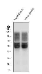
- Experimental details
- Western blot analysis of SLC2A1 using anti-SLC2A1 antibody (M00163-1). Electrophoresis was performed on a 5-20% SDS-PAGE gel at 70V (Stacking gel) / 90V (Resolving gel) for 2-3 hours. The sample well of each lane was loaded with 50ug of sample under reducing conditions. Lane 1: human placenta tissue lysates, Lane 2: human placenta tissue lysates. After Electrophoresis, proteins were transferred to a Nitrocellulose membrane at 150mA for 50-90 minutes. Blocked the membrane with 5% Non-fat Milk/ TBS for 1.5 hour at RT. The membrane was incubated with mouse anti-SLC2A1 antigen affinity purified monoclonal antibody (Catalog # M00163-1) at 0.5 μg/mL overnight at 4°C, then washed with TBS-0.1%Tween 3 times with 5 minutes each and probed with a goat anti-mouse IgG-HRP secondary antibody at a dilution of 1:10000 for 1.5 hour at RT. The signal is developed using an Enhanced Chemiluminescent detection (ECL) kit (Catalog # EK1001) with Tanon 5200 system. A specific band was detected for SLC2A1 at approximately 55KD. The expected band size for SLC2A1 is at 55KD.
- Additional image