Explore
 Validate
 Learn
LearnPA5-32428
antibody from Invitrogen Antibodies
Targeting: SLC2A1
CSE, DYT18, DYT9, GLUT, GLUT1, HTLVR
 Western blot
Antibody data
- Antibody Data
- Antigen structure
- References [1]
- Comments [0]
- Validations
- Western blot [2]
- Immunocytochemistry [1]
- Immunohistochemistry [1]
Submit
Validation data
Reference
Comment
Report error
- Product number
- PA5-32428 - Provider product page

- Provider
- Invitrogen Antibodies
- Product name
- GLUT1 Polyclonal Antibody
- Antibody type
- Polyclonal
- Antigen
- Synthetic peptide
- Description
- This antibody is predicted to react with bovine, chicken, mouse, porcine, rabbit and rat based on sequence homology.
- Concentration
- 0.4 mg/mL
Submitted references Arsenic trioxide disrupts glioma stem cells via promoting PML degradation to inhibit tumor growth.
Zhou W, Cheng L, Shi Y, Ke SQ, Huang Z, Fang X, Chu CW, Xie Q, Bian XW, Rich JN, Bao S
Oncotarget 2015 Nov 10;6(35):37300-15
Oncotarget 2015 Nov 10;6(35):37300-15
No comments: Submit comment
Supportive validation
- Submitted by
- Invitrogen Antibodies (provider)
- Main image

- Experimental details
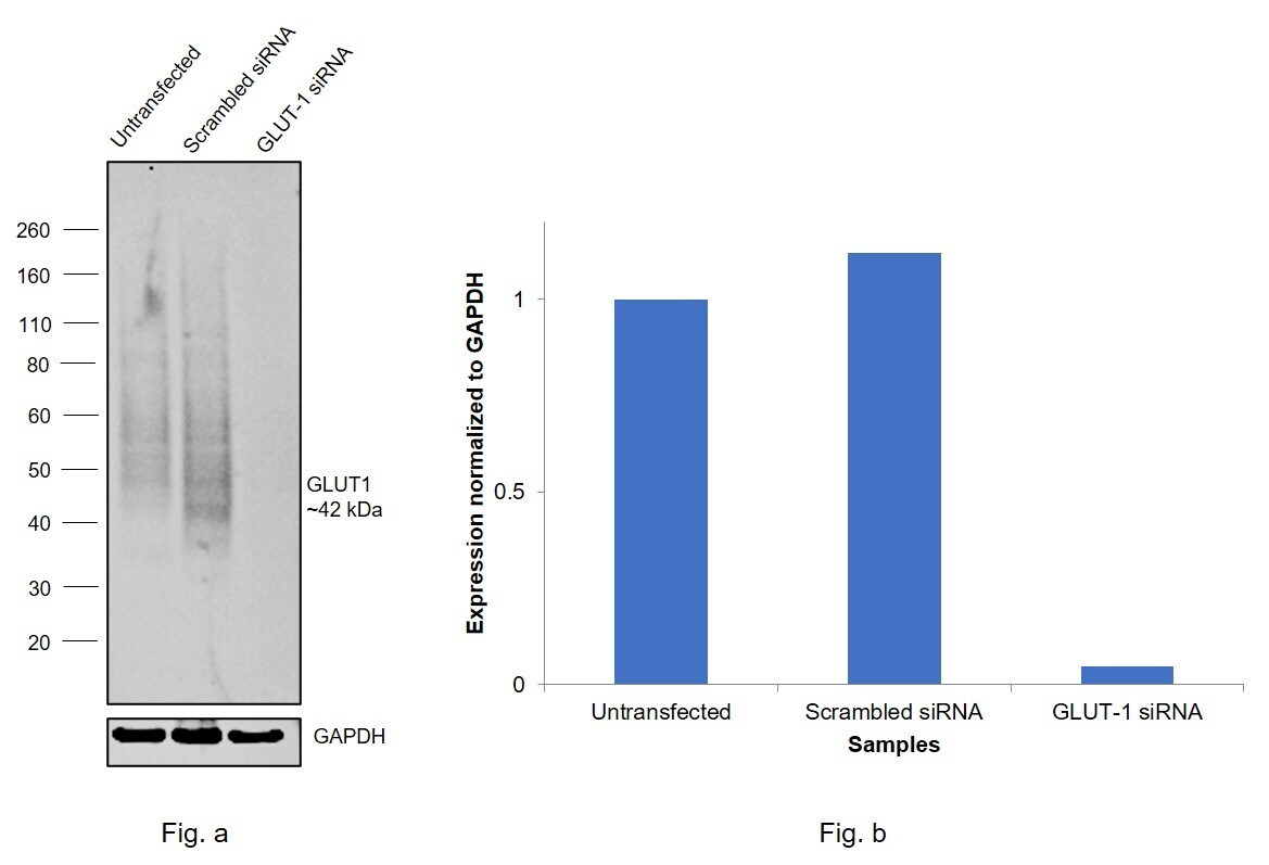
- Knockdown of GLUT1 was achieved by transfecting MCF7 with GLUT1 specific siRNAs (Silencer® select Product # s12927, s12926). Western blot analysis (Fig. a) was performed using Membrane enriched extracts from the GLUT1 knockdown cells (lane 3), non-targeting scrambled siRNA transfected cells (lane 2) and untransfected cells (lane 1). The blot was probed with GLUT1 Polyclonal Antibody (Product # PA5-32428, 0.5 µg/mL ) and Goat anti-Rabbit IgG (H+L) Superclonal™ Recombinant Secondary Antibody, HRP (Product # A27036, 1:4000). Densitometric analysis of this western blot is shown in histogram (Fig. b). Decrease in signal upon siRNA mediated knock down confirms that antibody is specific to GLUT1.The target GLUT-1 is known to show a streak like pattern in positive cell lines as seen in Lanes 1 and 2. Lane 3 shows almost complete loss of streak like pick up.
- Submitted by
- Invitrogen Antibodies (provider)
- Main image
- Experimental details
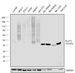
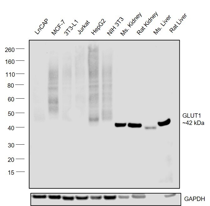
- Western blot was performed using Anti-GLUT1 Polyclonal Antibody (Product # PA5-32428) and a ~42kDa band corresponding to GLUT1 was observed across cell lines and tissues tested . Membrane enriched extracts (30 µg lysate) of LNCaP (Lane 1), MCF7 (Lane 2), 3T3-L1 (Lane 3), Jurkat (Lane 4), Hep G2 (Lane 5), NIH-3T3 (Lane 6), Mouse Kidney (Lane 7), Rat Kidney (Lane 8), Mouse Liver (Lane 9) and Rat Liver (Lane 10) were electrophoresed using NuPAGE™ 4-12% Bis-Tris Protein Gel (Product # NP0321BOX). Resolved proteins were then transferred onto a Nitrocellulose membrane (Product # LC2001) by iBlot® 2 Dry Blotting System (Product # IB21001). The blot was probed with the primary antibody (1:1000) and detected by chemiluminescence with Goat anti-Rabbit IgG (H+L) Superclonal™ Recombinant Secondary Antibody, HRP (Product # A27036, 1:4000) using the iBright FL 1000 (Product # A32752). Chemiluminescent detection was performed using Novex® ECL Chemiluminescent Substrate Reagent Kit (Product # WP20005). GLUT-1 shows a streak like signal in positive cell lines as seen in Lanes 1 to 6.
Supportive validation
- Submitted by
- Invitrogen Antibodies (provider)
- Main image
- Experimental details
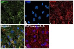
- Immunofluorescence analysis of GLUT1 was performed using 70% confluent log phase Hep G2 cells. The cells were fixed with 4% paraformaldehyde for 10 minutes, permeabilized with 0.1% Triton™ X-100 for 15 minutes, and blocked with 2% BSA for 45 minutes at room temperature. The cells were labeled with GLUT1 Polyclonal Antibody (Product # PA5-32428) at 1:200 in 0.1% BSA, incubated at 4 degree celsius overnight and then labeled with Donkey anti-Rabbit IgG (H+L) Highly Cross-Adsorbed Secondary Antibody, Alexa Fluor Plus 488 (Product # A32790), (1:2000), for 45 minutes at room temperature (Panel a: Blue). Nuclei (Panel b:Green) were stained with ProLong™ Diamond Antifade Mountant with DAPI (Product # P36962). F-actin (Panel c: Red) was stained with Rhodamine Phalloidin (Product # R415, 1:300). Panel d represents the merged image showing Plasma membrane and cytoplasm localization. Panel e represents control cells with no primary antibody to assess background. The images were captured at 60X magnification.
Supportive validation
- Submitted by
- Invitrogen Antibodies (provider)
- Main image
- Experimental details
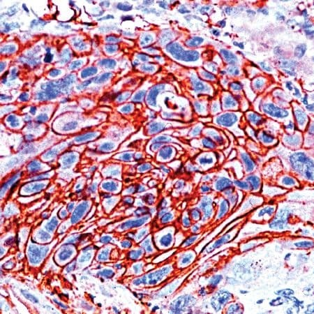
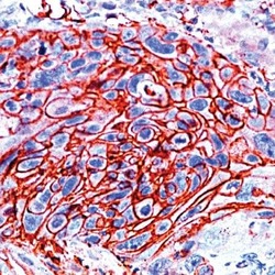
- Immunohistochemical analysis of GLUT-1 using anti-GLUT-1 Polyclonal Antibody (Product # PA5-32428) in Esophagus Cancer Tissue. The recommended dilution for this antibody in immunohistochemistry applications is 1:200.