 Explore
 Validate
 Learn
Learn Flow cytometry
Antibody data
- Antibody Data
- Antigen structure
- References [5]
- Comments [0]
- Validations
- Flow cytometry [2]
Submit
Validation data
Reference
Comment
Report error
- Product number
- FAB1418F - Provider product page

- Provider
- R&D Systems
- Product name
- Human Glut1 Fluorescein-conjugated Antibody
- Antibody type
- Monoclonal
- Description
- Protein A or G purified from hybridoma culture supernatant. Detects human Glut1. Stains human Glut1-transfected NS0 cells, but not NS0 control transfectants. Although Human Glut1 Antibody detects Glut1 on the surface of T cells (1, 2), it does not detect it on erythrocytes (5). The reason for this discrepancy is not understood, but may be related to conformational or post-translational modification differences.
- Reactivity
- Human
- Host
- Mouse
- Antigen sequence
AAA52571- Isotype
- IgG
- Antibody clone number
- 202915
- Vial size
- 100 Tests
- Storage
- Protect from light. Do not freeze. 12 months from date of receipt, 2 to 8 °C as supplied.
Submitted references Immunometabolic and Lipidomic Markers Associated With the Frailty Index and Quality of Life in Aging HIV+ Men on Antiretroviral Therapy.
PD-1 alters T-cell metabolic reprogramming by inhibiting glycolysis and promoting lipolysis and fatty acid oxidation.
Separate cellular localizations of human T-lymphotropic virus 1 (HTLV-1) Env and glucose transporter type 1 (GLUT1) are required for HTLV-1 Env-mediated fusion and infection.
Glucose transporter 1-positive endothelial cells in infantile hemangioma exhibit features of facultative stem cells.
Increased glucose metabolic activity is associated with CD4+ T-cell activation and depletion during chronic HIV infection.
Yeoh HL, Cheng AC, Cherry CL, Weir JM, Meikle PJ, Hoy JF, Crowe SM, Palmer CS
EBioMedicine 2017 Aug;22:112-121
EBioMedicine 2017 Aug;22:112-121
PD-1 alters T-cell metabolic reprogramming by inhibiting glycolysis and promoting lipolysis and fatty acid oxidation.
Patsoukis N, Bardhan K, Chatterjee P, Sari D, Liu B, Bell LN, Karoly ED, Freeman GJ, Petkova V, Seth P, Li L, Boussiotis VA
Nature communications 2015 Mar 26;6:6692
Nature communications 2015 Mar 26;6:6692
Separate cellular localizations of human T-lymphotropic virus 1 (HTLV-1) Env and glucose transporter type 1 (GLUT1) are required for HTLV-1 Env-mediated fusion and infection.
Maeda Y, Terasawa H, Tanaka Y, Mitsuura C, Nakashima K, Yusa K, Harada S
Journal of virology 2015 Jan;89(1):502-11
Journal of virology 2015 Jan;89(1):502-11
Glucose transporter 1-positive endothelial cells in infantile hemangioma exhibit features of facultative stem cells.
Huang L, Nakayama H, Klagsbrun M, Mulliken JB, Bischoff J
Stem cells (Dayton, Ohio) 2015 Jan;33(1):133-45
Stem cells (Dayton, Ohio) 2015 Jan;33(1):133-45
Increased glucose metabolic activity is associated with CD4+ T-cell activation and depletion during chronic HIV infection.
Palmer CS, Ostrowski M, Gouillou M, Tsai L, Yu D, Zhou J, Henstridge DC, Maisa A, Hearps AC, Lewin SR, Landay A, Jaworowski A, McCune JM, Crowe SM
AIDS (London, England) 2014 Jan 28;28(3):297-309
AIDS (London, England) 2014 Jan 28;28(3):297-309
No comments: Submit comment
Supportive validation
- Submitted by
- R&D Systems (provider)
- Main image
- Experimental details
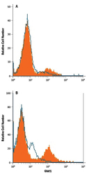
- Detection of Glut1 in Jurkat Human Cell Line by Flow Cytometry. Jurkat human acute T cell leukemia cell line either (A) untreated or (B) cultured in nutrient-depleted media was stained with Mouse Anti-Human Glut1 Fluorescein-conjugated Monoclonal Antibody (Catalog # FAB1418F, filled histogram) or isotype control antibody (Catalog # IC0041F, open histogram). View our protocol for Staining Membrane-associated Proteins.

- Submitted by
- R&D Systems (provider)
- Main image
- Experimental details
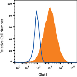
- Detection of Glut1 in HepG2 Human Cell Line by Flow Cytometry. HepG2 human hepatocellular carcinoma cell line was stained with Mouse Anti-Human Glut1 Fluorescein-conjugated Monoclonal Antibody (Catalog # FAB1418F, filled histogram) or isotype control antibody (Catalog # IC0041F, open histogram). View our protocol for Staining Membrane-associated Proteins.
