Explore
 Validate
 Learn
Learn Western blot
 Immunocytochemistry
Antibody data
- Antibody Data
- Antigen structure
- References [1]
- Comments [0]
- Validations
- Western blot [9]
- ELISA [1]
- Immunoprecipitation [2]
- Immunohistochemistry [2]
- Flow cytometry [1]
Submit
Validation data
Reference
Comment
Report error
- Product number
- NBP1-33197 - Provider product page

- Provider
- Novus Biologicals
- Proper citation
- Novus Cat#NBP1-33197, RRID:AB_2279149
- Product name
- Rabbit Polyclonal Glypican 1 Antibody
- Antibody type
- Polyclonal
- Description
- Immunogen affinity purified.
- Reactivity
- Human, Mouse, Rat
- Host
- Rabbit
- Isotype
- IgG
- Vial size
- 0.1 ml
- Storage
- Aliquot and store at -20C or -80C. Avoid freeze-thaw cycles.
Submitted references Ginsenoside Rg1 attenuates high glucose-induced endothelial barrier dysfunction in human umbilical vein endothelial cells by protecting the endothelial glycocalyx.
Zhu T, Wang H, Wang L, Zhong X, Huang W, Deng X, Guo H, Xiong J, Xu Y, Fan J
Experimental and therapeutic medicine 2019 May;17(5):3727-3733
Experimental and therapeutic medicine 2019 May;17(5):3727-3733
No comments: Submit comment
Supportive validation
- Submitted by
- Novus Biologicals (provider)
- Main image
- Experimental details
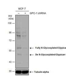
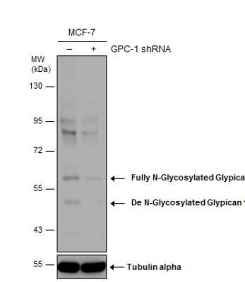
- Western Blot: Glypican 1 Antibody [NBP1-33197] - Non-transfected (-) and transfected (+) MCF-7 whole cell extracts (50 ug) were separated by 7.5% SDS-PAGE, and the membrane was blotted with Glypican 1 antibody [N3C3].The observed M.W. is based on the publication: PMID: 22351761, 26203194 and 21932778
- Submitted by
- Novus Biologicals (provider)
- Main image
- Experimental details
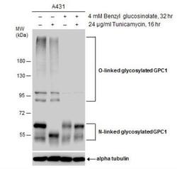
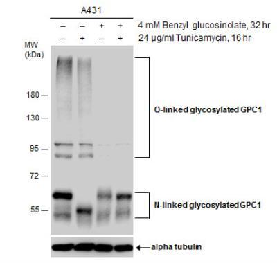
- Western Blot: Glypican 1 Antibody [NBP1-33197] - Untreated (-) and treated (+) A431 whole cell extracts (30 ug) were separated by 7.5% SDS-PAGE, and the membrane was blotted with Glypican 1 antibody [N3C3]. The HRP-conjugated anti-rabbit IgG antibody was used to detect the primary antibody.
- Submitted by
- Novus Biologicals (provider)
- Main image
- Experimental details
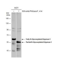
- Western Blot: Glypican 1 Antibody [NBP1-33197] - Untreated (-) and treated (+) A431 whole cell extracts (30 ug) were separated by 7.5% SDS-PAGE, and the membrane was blotted with Glypican 1 antibody [N3C3].The observed M.W. is based on the following publications: PMID: 22351761, 26203194 and 21932778

- Submitted by
- Novus Biologicals (provider)
- Main image
- Experimental details
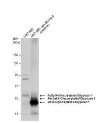
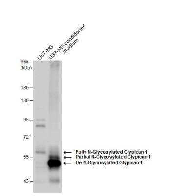
- Western Blot: Glypican 1 Antibody [NBP1-33197] - U87-MG whole cell extract and conditioned medium (30 ug) were separated by 7.5% SDS-PAGE, and the membrane was blotted with Glypican 1 antibody [N3C3] diluted at 1:1500. The HRP-conjugated anti-rabbit IgG antibody ] was used to detect the primary antibody.
- Submitted by
- Novus Biologicals (provider)
- Main image
- Experimental details
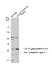
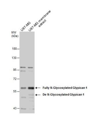
- Western Blot: Glypican 1 Antibody [NBP1-33197] - U87-MG whole cell and membrane extracts (30 ug) were separated by 7.5% SDS-PAGE, and the membrane was blotted with Glypican 1 antibody [N3C3] diluted at 1:1500. The HRP-conjugated anti-rabbit IgG antibody was used to detect the primary antibody.
- Submitted by
- Novus Biologicals (provider)
- Main image
- Experimental details
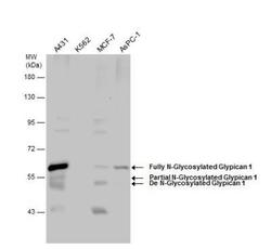
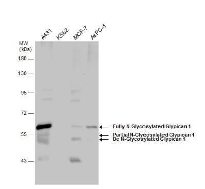
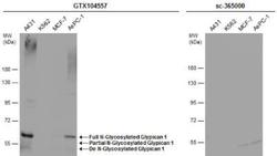
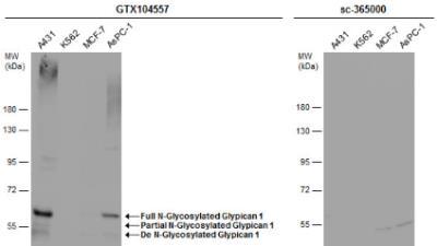
- Western Blot: Glypican 1 Antibody [NBP1-33197] - Various whole cell extracts (30 ug) were separated by 7.5% SDS-PAGE, and the membrane was blotted with Glypican 1 antibody [N3C3] diluted at 1:1500.The observed M.W. is based on the publication: PMID: 22351761, 26203194 and 21932778
- Submitted by
- Novus Biologicals (provider)
- Main image
- Experimental details
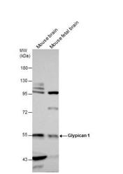
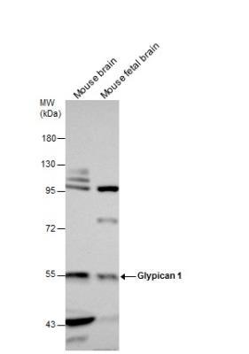
- Western Blot: Glypican 1 Antibody [NBP1-33197] - Mouse tissue extracts (50 ug) were separated by 7.5% SDS-PAGE, and the membrane was blotted with Glypican 1 antibody [N3C3] diluted at 1:1500.
- Submitted by
- Novus Biologicals (provider)
- Main image
- Experimental details
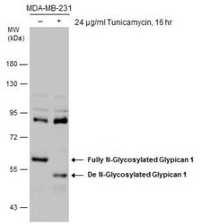
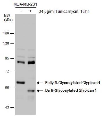
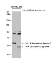
- Western Blot: Glypican 1 Antibody [NBP1-33197] - Untreated (-) and treated (+) MDA-MB-231 whole cell extracts (30 ug) were separated by 7.5% SDS-PAGE, and the membrane was blotted with Glypican 1 antibody [N3C3]. HRP-conjugated anti-rabbit IgG antibody was used to detect the primary antibody. The observed M.W. is based on the following publications: PMID: 22351761, 26203194 and 21932778
- Submitted by
- Novus Biologicals (provider)
- Main image
- Experimental details
- Western Blot: Glypican 1 Antibody [NBP1-33197] - Various whole cell extracts (30 ug) were separated by 7.5% SDS-PAGE, and the membranes were blotted with Glypican 1 antibody [N3C3] diluted at 1:500 and competitor's antibody (sc-365000) diluted at 1:500. The HRP-conjugated anti-rabbit IgG antibody was used to detect the primary antibody.
Supportive validation
- Submitted by
- Novus Biologicals (provider)
- Main image
- Experimental details
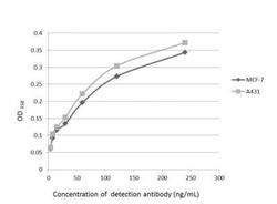
- ELISA: Glypican 1 Antibody [NBP1-33197] - An ELISA plate is coated with MCF-7 and A431 cells. The coated cells are detected with Glypican 1 antibody at concentration ranged from 7.5 to 240 ng/mL.

Supportive validation
- Submitted by
- Novus Biologicals (provider)
- Main image
- Experimental details
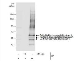
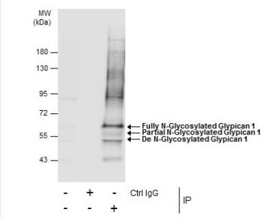
- Immunoprecipitation: Glypican 1 Antibody [NBP1-33197] - Untreated (-) and treated (+) MDA-MB-231 whole cell extracts (30 ug) were separated by 7.5% SDS-PAGE, and the membrane was blotted with Glypican 1 antibody [N3C3] diluted at 1:1500.The observed M.W. is based on the publication: PMID: 22351761, 26203194 and 21932778

- Submitted by
- Novus Biologicals (provider)
- Main image
- Experimental details
- Immunoprecipitation: Glypican 1 Antibody [NBP1-33197] - Immunoprecipitation of Glypican 1 protein from MCF-7 whole cell extracts using 5 ug of Glypican 1 antibody [N3C3].
Supportive validation
- Submitted by
- Novus Biologicals (provider)
- Main image

- Experimental details
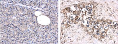
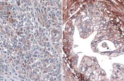
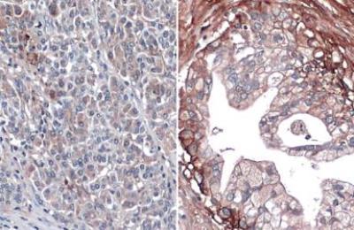
- Immunohistochemistry-Paraffin: Glypican 1 Antibody [NBP1-33197] - Analysis of paraffin-embedded human normal pancreas (left) or pancreatic adenocarcinoma (grade II) (right) tissues using Glypican-1 antibody [N3C3] at a 1:250 dilution.
- Submitted by
- Novus Biologicals (provider)
- Main image
- Experimental details
- Immunohistochemistry-Paraffin: Glypican 1 Antibody [NBP1-33197] - Paraffin-embedded human normal pancreas (left) and pancreatic cancer (right). Glypican 1 stained by Glypican 1 antibody [N3C3] diluted at 1:500. Antigen Retrieval: Citrate buffer, pH 6.0, 15 min
Supportive validation
- Submitted by
- Novus Biologicals (provider)
- Main image

- Experimental details
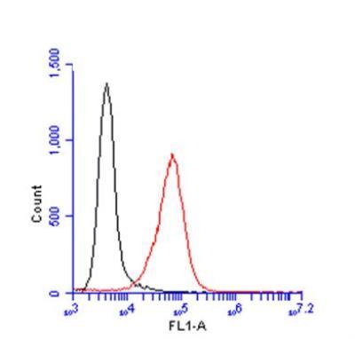
- Flow Cytometry: Glypican 1 Antibody [NBP1-33197] - Sample: A431 cell. Black: Unlabelled sample was used as a control. Red: Glypican 1 antibody [N3C3]dilution: 1:100. Acquisition of 20,000 events were collected using a Dylight 488-conjugated secondary antibody for FACS analysis.