Explore
 Validate
 Learn
Learn Western blot
 Immunohistochemistry
Antibody data
- Antibody Data
- Antigen structure
- References [2]
- Comments [0]
- Validations
- Immunohistochemistry [1]
- Other assay [1]
Submit
Validation data
Reference
Comment
Report error
- Product number
- PA5-23237 - Provider product page

- Provider
- Invitrogen Antibodies
- Product name
- GPR35 Polyclonal Antibody
- Antibody type
- Polyclonal
- Antigen
- Other
- Reactivity
- Human, Mouse
- Host
- Rabbit
- Isotype
- IgG
- Vial size
- 100 μg
- Concentration
- 1 mg/mL
- Storage
- Store at 4°C short term. For long term storage, store at -20°C, avoiding freeze/thaw cycles.
Submitted references Inhibition of GPR35 Preserves Mitochondrial Function After Myocardial Infarction by Targeting Calpain 1/2.
Activation of GPR35 protects against cerebral ischemia by recruiting monocyte-derived macrophages.
Chen K, He L, Li Y, Li X, Qiu C, Pei H, Yang D
Journal of cardiovascular pharmacology 2020 Jun;75(6):556-563
Journal of cardiovascular pharmacology 2020 Jun;75(6):556-563
Activation of GPR35 protects against cerebral ischemia by recruiting monocyte-derived macrophages.
Sharmin O, Abir AH, Potol A, Alam M, Banik J, Rahman AFMT, Tarannum N, Wadud R, Habib ZF, Rahman M
Scientific reports 2020 Jun 10;10(1):9400
Scientific reports 2020 Jun 10;10(1):9400
No comments: Submit comment
Supportive validation
- Submitted by
- Invitrogen Antibodies (provider)
- Main image
- Experimental details
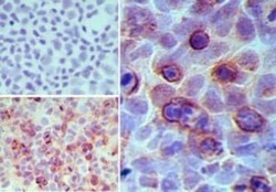
- Immunohistochemical analysis of GPR35 in paraffin-embedded formalin-fixed MCF7 cells. Samples were incubated in GPR35 polyclonal antibody (Product # PA5-23237) using a dilution of 5 µg/mL. Isotype control (top left) and this antibody (bottom left, right).

Supportive validation
- Submitted by
- Invitrogen Antibodies (provider)
- Main image
- Experimental details
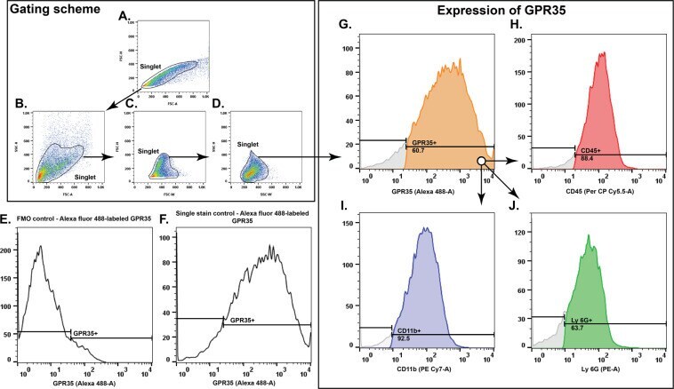
- Figure 3 Cellular expression of GPR35 in the ischemic brain. ( A-D ) Represents the gating scheme for doublet discrimination and isolation of the singlet population. ( E ) Fluorescence minus one (FMO) control for Alexa fluor 488 labeled GPR35 positive cells. ( F ) Single stain control for Alexa fluor 488 labeled GPR35 positive cells. ( G-J ) Representation of the flowcytometry analysis revealing that the CD45 ( H ), CD11b ( I ), and Ly-6G ( J ) positive cells express GPR35 ( G ).