Explore
 Validate
 Learn
Learn Western blot
 Other assay
Antibody data
- Antibody Data
- Antigen structure
- References [2]
- Comments [0]
- Validations
- Other assay [1]
Submit
Validation data
Reference
Comment
Report error
- Product number
- PA1-059 - Provider product page

- Provider
- Invitrogen Antibodies
- Product name
- Adiponectin Receptor 1 Polyclonal Antibody
- Antibody type
- Polyclonal
- Antigen
- Synthetic peptide
- Description
- PA1-059 detects human and rat adiponectin receptor 1. PA1-059 has been successfully used in Western blot procedures. By Western blot, this antibody detects an ~42 kDa protein corresponding to human adiponectin receptor 1 from transfected hepatocytes. The PA1-059 immunogen is a synthetic peptide corresponding to residues R(20) E A D T V E L A E L G P L L E E K(37) of human Adiponectin Receptor 1.
- Reactivity
- Human, Rat
- Host
- Rabbit
- Isotype
- IgG
- Vial size
- 100 μg
- Concentration
- 1 mg/mL
- Storage
- -20°C, Avoid Freeze/Thaw Cycles
Submitted references Adiponectin receptor agonists inhibit leptin induced pSTAT3 and in vivo pancreatic tumor growth.
Increase of adiponectin receptor gene expression by physical exercise in soleus muscle of obese Zucker rats.
Messaggio F, Mendonsa AM, Castellanos J, Nagathihalli NS, Gorden L, Merchant NB, VanSaun MN
Oncotarget 2017 Oct 17;8(49):85378-85391
Oncotarget 2017 Oct 17;8(49):85378-85391
Increase of adiponectin receptor gene expression by physical exercise in soleus muscle of obese Zucker rats.
Chang SP, Chen YH, Chang WC, Liu IM, Cheng JT
European journal of applied physiology 2006 May;97(2):189-95
European journal of applied physiology 2006 May;97(2):189-95
No comments: Submit comment
Supportive validation
- Submitted by
- Invitrogen Antibodies (provider)
- Main image

- Experimental details
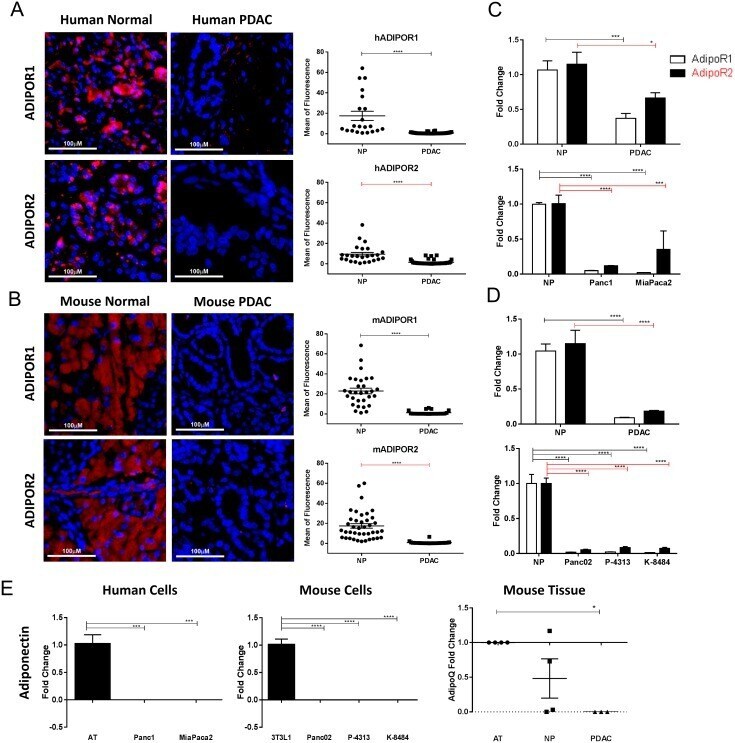
- Figure 1 Adiponectin receptor levels are reduced in pancreatic cancer Human and murine normal and pancreatic cancer samples were immunostained for ADIPOR1 and ADIPOR2. (A) strong staining is present in the acinar tissue of human normal samples for both receptors but is decreased in the human pancreatic tumor samples. (B) murine pancreatic cancer tissue samples from PKT mice demonstrate a high level of staining in the adjacent acinar tissue and a reduced staining in the dysplastic pancreatic tumor. Graphs represent quantitative analysis of mean fluorescence from single channel images. Scale bar is 100mum. (C-D) quantitative real time PCR analysis was used to determine the relative expression of AdipoR1 (white bars) and AdipoR2 (black bars) in human and murine pancreatic tumor samples (PDAC) as well as in pancreatic cancer cell lines, relative to the level expressed in the normal pancreas (NP) tissue. (E) quantitative real time PCR analysis for adiponectin expression in human and mouse pancreatic cancer cell lines and mouse tissue, relative to human adipose tissue (AT), 3T3L1 cells differentiated to adipocytes (3T3L1) and mouse AT, respectively. Statistical analysis was performed separately for ADIPOR1 and ADIPOR2 protein levels (unpaired t-test) as well as AdipoR1 and AdipoR2 expression (two-way Anova), or adiponectin expression (ordinary one-way Anova) (*P