Explore
 Validate
 Learn
Learn Western blot
Antibody data
- Antibody Data
- Antigen structure
- References [0]
- Comments [0]
- Validations
- Western blot [2]
- ELISA [2]
- Immunocytochemistry [4]
- Flow cytometry [2]
- Other assay [1]
Submit
Validation data
Reference
Comment
Report error
- Product number
- 710340 - Provider product page

- Provider
- Invitrogen Antibodies
- Product name
- TIMP4 Recombinant Superclonal™ Antibody (13HCLC)
- Antibody type
- Other
- Antigen
- Other
- Description
- Recombinant rabbit Superclonal™ antibodies are unique offerings from Thermo Fisher Scientific. They are comprised of a selection of multiple different recombinant monoclonal antibodies, providing the best of both worlds - the sensitivity of polyclonal antibodies with the specificity of monoclonal antibodies - all delivered with the consistency only found in a recombinant antibody. While functionally the same as a polyclonal antibody - recognizing multiple epitope sites on the target and producing higher detection sensitivity for low abundance targets - a recombinant rabbit Superclonal™ antibody has a known mixture of light and heavy chains. The exact population can be produced in every lot, circumventing the biological variability typically associated with polyclonal antibody production. Note: Formerly called “Recombinant polyclonal antibody”, this product is now rebranded as “Recombinant Superclonal™ antibody”. The physical product and the performance remain unchanged.
- Reactivity
- Human
- Host
- Rabbit
- Isotype
- IgG
- Antibody clone number
- 13HCLC
- Vial size
- 100 μg
- Concentration
- 0.5 mg/mL
- Storage
- Store at 4°C short term. For long term storage, store at -20°C, avoiding freeze/thaw cycles.
No comments: Submit comment
Supportive validation
- Submitted by
- Invitrogen Antibodies (provider)
- Main image
- Experimental details
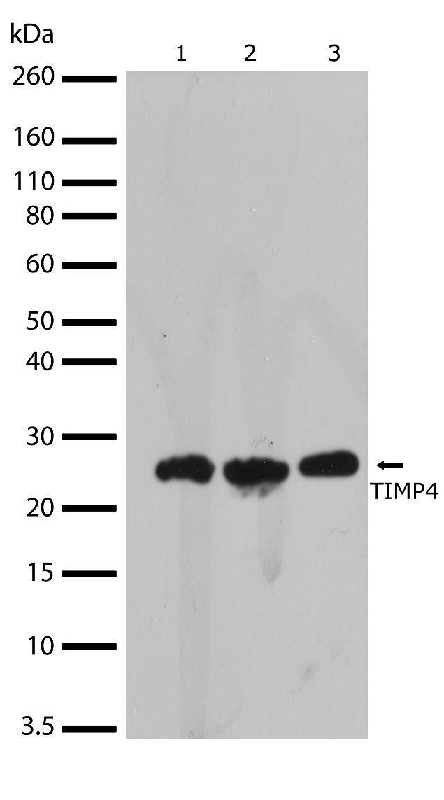
- Western blot analysis of TIMP4 in HeLa, HEK293 and MDA-MB-231 whole cell extracts using a TIMP4 Recombinant Rabbit Superclonal™ Antibody (Product # 710340) at a dilution of 2 µg/mL. Samples were detected using chemiluminescence (ECL). Results show a band at ~25kDa.
- Submitted by
- Invitrogen Antibodies (provider)
- Main image
- Experimental details
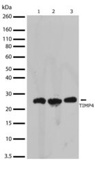
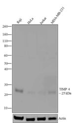
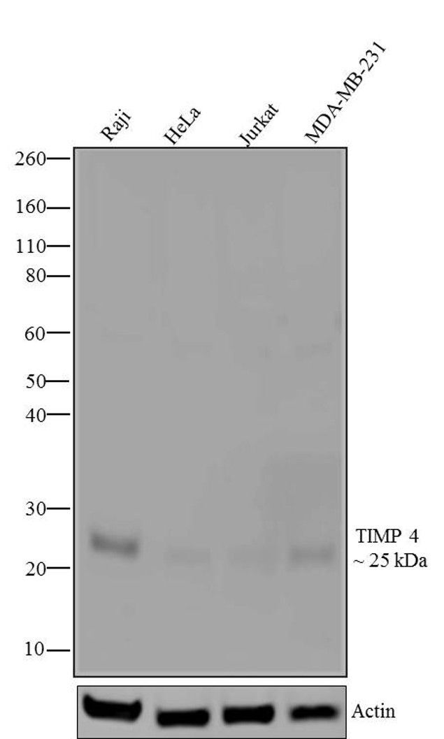
- Western blot analysis of TIMP4was performed by loading 20 µg of Raji (lane1), HeLa (lane2), Jurkat (lane3) and MDA-MB-231 (lane4) cell lysates using Novex®NuPAGE®4-12 % Bis-Tris gel (Product # NP0321BOX), XCell SureLock Electrophoresis System (Product # EI0002), Novex® Sharp Pre-Stained Protein Standard (Product # LC5800), and iBlot® Dry Blotting System (Product # IB21001). Proteins were transferred to a nitrocellulose membrane and blocked with 5 % skim milk for 1 hour at room temperature. TIMP4was detected at ~25 kDa using TIMP4 Recombinant Rabbit Superclonal™ Antibody (Product # 710340) at 1 µg-3 µg/mL in 2.5 % skim milk at 4°C overnight on a rocking platform. Goat anti-Rabbit IgG-HRP Secondary Antibody (Product # G-21234) at 1:5000 dilution was used and chemiluminescent detection was performed using Pierce™ ECL Western blotting Substrate (Product # 32106).
Supportive validation
- Submitted by
- Invitrogen Antibodies (provider)
- Main image
- Experimental details
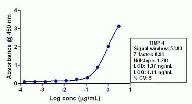
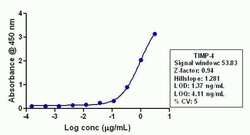
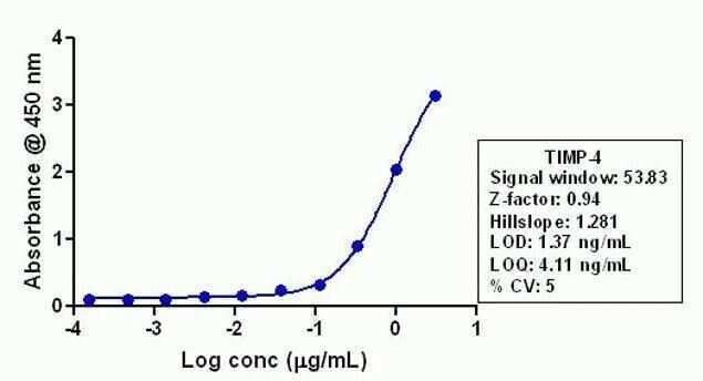
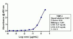
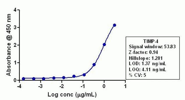
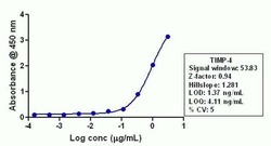
- Indirect ELISA analysis of endogenous TIMP4 HeLa lysate coated onto the plate using a TIMP4 Recombinant Rabbit Polyclonal Antibody (Product # 710340) at various dilutions. A non-linear regression analysis was performed (4 PL) and LOD and LOQ for the antibody were determined.
- Submitted by
- Invitrogen Antibodies (provider)
- Main image
- Experimental details
- Indirect ELISA analysis of endogenous TIMP4 HeLa lysate coated onto the plate using a TIMP4 Recombinant Rabbit Superclonal™ Antibody (Product # 710340) at various dilutions. A non-linear regression analysis was performed (4 PL) and LOD and LOQ for the antibody were determined.
Supportive validation
- Submitted by
- Invitrogen Antibodies (provider)
- Main image
- Experimental details
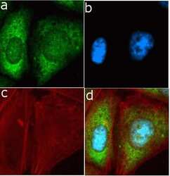
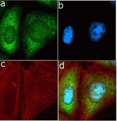
- Immunofluorescent analysis of TIMP4 in U2OS cells using a TIMP4 Recombinant Rabbit Polyclonal Antibody (Product # 710340) followed by detection using an Alexa Fluor 488-conjugated Goat anti-Rabbit secondary antibody (green) (Image A). Nuclei were stained using DAPI (Image B) and actin stained with Alexa Fluor 594 phalloidin (red) (image C). Image D is a composite image showing cytoplasmic localization of TIMP4.

- Submitted by
- Invitrogen Antibodies (provider)
- Main image
- Experimental details
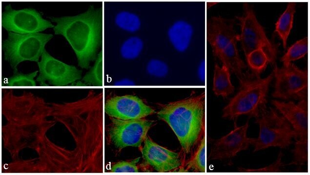
- Immunofluorescent analysis of TIMP4 was done on 70% confluent log phase HeLa cells. The cells were fixed with 4% paraformaldehyde for 15 minutes; permeabilized with 0.25% Triton X-100 for 10 minutes followed by blocking with 5% BSA for 1 hour at room temperature. The cells were incubated with TIMP4 Recombinant Rabbit Polyclonal Antibody (Product # 710340) at 1 µg-2 µg in 1% BSA and incubated for 3 hours at room temperature and then labeled with Alexa Fluor® 488 Goat anti-Rabbit IgG Secondary Antibody (Product # A-11008) at a dilution of 1:400 for 30 minutes at room temperature (Panel a: green). Nuclei (Panel b: blue) were stained with SlowFade® Gold Antifade Mountant with DAPI (Product # S36938). F-actin (Panel c: red) was stained with Alexa Fluor® 594 Phalloidin (Product # A12381). Panel d is a merged image showing cytoplasmic localization of TIMP4. Panel e shows no primary antibody control. The images were captured at 20X magnification.

- Submitted by
- Invitrogen Antibodies (provider)
- Main image
- Experimental details
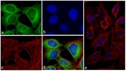
- Immunofluorescent analysis of TIMP4 in U2OS cells using a TIMP4 Recombinant Rabbit Superclonal™ Antibody (Product # 710340) followed by detection using an Alexa Fluor 488-conjugated Goat anti-Rabbit secondary antibody (green) (Image A). Nuclei were stained using DAPI (Image B) and actin stained with Alexa Fluor 594 phalloidin (red) (image C). Image D is a composite image showing cytoplasmic localization of TIMP4.

- Submitted by
- Invitrogen Antibodies (provider)
- Main image

- Experimental details
- Immunofluorescent analysis of TIMP4 was done on 70% confluent log phase HeLa cells. The cells were fixed with 4% paraformaldehyde for 15 minutes; permeabilized with 0.25% Triton X-100 for 10 minutes followed by blocking with 5% BSA for 1 hour at room temperature. The cells were incubated with TIMP4 Recombinant Rabbit Superclonal™ Antibody (Product # 710340) at 1 µg-2 µg in 1% BSA and incubated for 3 hours at room temperature and then labeled with Alexa Fluor® 488 Goat anti-Rabbit IgG Secondary Antibody (Product # A-11008) at a dilution of 1:400 for 30 minutes at room temperature (Panel a: green). Nuclei (Panel b: blue) were stained with SlowFade® Gold Antifade Mountant with DAPI (Product # S36938). F-actin (Panel c: red) was stained with Alexa Fluor® 594 Phalloidin (Product # A12381). Panel d is a merged image showing cytoplasmic localization of TIMP4. Panel e shows no primary antibody control. The images were captured at 20X magnification.
Supportive validation
- Submitted by
- Invitrogen Antibodies (provider)
- Main image
- Experimental details
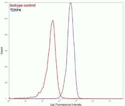
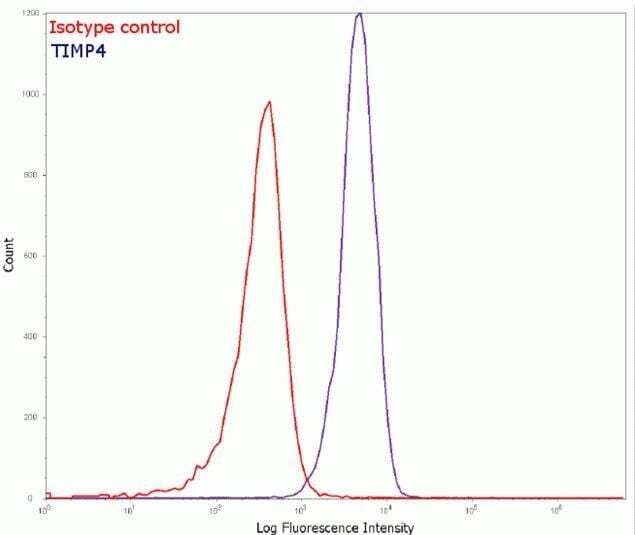
- Flow cytometry analysis of TIMP4 in Hela cells using a TIMP4 Recombinant Rabbit Polyclonal Antibody (Product # 710340). Cells were fixed and permeabilized using FIX & PERM (Product # GAS-004) reagent, and detection was performed using an Alexa Fluor 488 Goat anti-Rabbit IgG (right peak) compared to an isotype control (left peak).
- Submitted by
- Invitrogen Antibodies (provider)
- Main image

- Experimental details
- Flow cytometry analysis of TIMP4 in Hela cells using a TIMP4 Recombinant Rabbit Superclonal™ Antibody (Product # 710340). Cells were fixed and permeabilized using FIX & PERM (Product # GAS-004) reagent, and detection was performed using an Alexa Fluor 488 Goat anti-Rabbit IgG (right peak) compared to an isotype control (left peak).

Supportive validation
- Submitted by
- Invitrogen Antibodies (provider)
- Main image
- Experimental details
- Indirect ELISA analysis of endogenous TIMP4 HeLa lysate coated onto the plate using a TIMP4 Recombinant Rabbit Polyclonal Antibody (Product # 710340) at various dilutions. A non-linear regression analysis was performed (4 PL) and LOD and LOQ for the antibody were determined.