 Explore
 Validate
 Learn
Learn Western blot
 Immunohistochemistry
Antibody data
- Antibody Data
- Antigen structure
- References [0]
- Comments [0]
- Validations
- Immunohistochemistry [5]
Submit
Validation data
Reference
Comment
Report error
- Product number
- LS-C744845 - Provider product page

- Provider
- LSBio
- Product name
- TIMP4 Antibody (clone 9:4-7) LS-C744845
- Antibody type
- Monoclonal
- Description
- Protein A affinity chromatography
- Reactivity
- Human, Mouse
- Host
- Mouse
- Isotype
- IgG
- Antibody clone number
- 9:4-7
- Storage
- Store vial at -20°C or below prior to opening. Dilute 1:10 to minimize loss. Store the vial at -20°C or below after dilution. Avoid freeze-thaw cycles.
No comments: Submit comment
Supportive validation
- Submitted by
- LSBio (provider)
- Enhanced method
- Genetic validation
- Main image

- Experimental details
- Immunohistochemistry of Mouse anti-TMIP4 antibody. Tissue: kidney. Fixation: formalin fixed paraffin embedded. Antigen retrieval: not required. Primary antibody: anti-TMIP4 antibody at 10 µg/mL for 1 h at RT. Secondary antibody: Peroxidase mouse secondary antibody at 1:10,000 for 45 min at RT. Staining: TMIP4 as precipitated red signal with hematoxylin purple nuclear counterstain.

- Submitted by
- LSBio (provider)
- Enhanced method
- Genetic validation
- Main image

- Experimental details
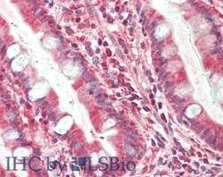
- Immunohistochemistry of Mouse anti-TIMP4 antibody. Tissue: small intestine. Fixation: formalin fixed paraffin embedded. Antigen retrieval: not required. Primary antibody: anti-TIMP4 antibody at 10 µg/mL for 1 h at RT. Secondary antibody: Peroxidase mouse secondary antibody at 1:10,000 for 45 min at RT. Staining: TIMP4 as precipitated red signal with hematoxylin purple nuclear counterstain.

- Submitted by
- LSBio (provider)
- Main image

- Experimental details
- Immunohistochemistry of Mouse anti-TMIP4 antibody. Tissue: kidney. Fixation: formalin fixed paraffin embedded. Antigen retrieval: not required. Primary antibody: anti-TMIP4 antibody at 10 µg/mL for 1 h at RT. Secondary antibody: Peroxidase mouse secondary antibody at 1:10,000 for 45 min at RT. Staining: TMIP4 as precipitated red signal with hematoxylin purple nuclear counterstain.

- Submitted by
- LSBio (provider)
- Main image
- Experimental details
- Immunohistochemistry of Mouse anti-TIMP4 antibody. Tissue: small intestine. Fixation: formalin fixed paraffin embedded. Antigen retrieval: not required. Primary antibody: anti-TIMP4 antibody at 10 µg/mL for 1 h at RT. Secondary antibody: Peroxidase mouse secondary antibody at 1:10,000 for 45 min at RT. Staining: TIMP4 as precipitated red signal with hematoxylin purple nuclear counterstain.

- Submitted by
- LSBio (provider)
- Main image

- Experimental details
- Immunohistochemistry of Mouse Anti-TIMP4 Antibody. Tissue: human breast carcinoma. Fixation: formalin fixed paraffin embedded. Antigen retrieval: not required. Primary antibody: a) isotype specific IgG2bk monoclonal; b) TIMP-4 antibody; and c) TIMP-4 each at 10 µg/mL. Secondary antibody: Peroxidase mouse secondary antibody at 1:10,000 for 45 min at RT. Localization: TIMP-4 is secreted. Staining: TIMP-4 as precipitated brown signal.
