Explore
 Validate
 Learn
Learn Western blot
 Immunocytochemistry
Antibody data
- Antibody Data
- Antigen structure
- References [6]
- Comments [0]
- Validations
- Immunocytochemistry [6]
- Immunohistochemistry [1]
- Other assay [5]
Submit
Validation data
Reference
Comment
Report error
- Product number
- PA1-093X - Provider product page

- Provider
- Invitrogen Antibodies
- Product name
- ARF6 Polyclonal Antibody
- Antibody type
- Polyclonal
- Antigen
- Recombinant protein fragment
- Reactivity
- Human, Mouse
- Host
- Rabbit
- Isotype
- IgG
- Vial size
- 20 μL
- Concentration
- 1 mg/mL
- Storage
- -20°C, Avoid Freeze/Thaw Cycles
Submitted references SARS-CoV-2 pseudovirus enters the host cells through spike protein-CD147 in an Arf6-dependent manner.
A mosquito salivary protein promotes flavivirus transmission by activation of autophagy.
Heterozygous loss of function of IQSEC2/Iqsec2 leads to increased activated Arf6 and severe neurocognitive seizure phenotype in females.
Syndecan 1 is a critical mediator of macropinocytosis in pancreatic cancer.
Which Way In? The RalF Arf-GEF Orchestrates Rickettsia Host Cell Invasion.
Secretion of soluble vascular endothelial growth factor receptor 1 (sVEGFR1/sFlt1) requires Arf1, Arf6, and Rab11 GTPases.
Zhou YQ, Wang K, Wang XY, Cui HY, Zhao Y, Zhu P, Chen ZN
Emerging microbes & infections 2022 Dec;11(1):1135-1144
Emerging microbes & infections 2022 Dec;11(1):1135-1144
A mosquito salivary protein promotes flavivirus transmission by activation of autophagy.
Sun P, Nie K, Zhu Y, Liu Y, Wu P, Liu Z, Du S, Fan H, Chen CH, Zhang R, Wang P, Cheng G
Nature communications 2020 Jan 14;11(1):260
Nature communications 2020 Jan 14;11(1):260
Heterozygous loss of function of IQSEC2/Iqsec2 leads to increased activated Arf6 and severe neurocognitive seizure phenotype in females.
Jackson MR, Loring KE, Homan CC, Thai MH, Määttänen L, Arvio M, Jarvela I, Shaw M, Gardner A, Gecz J, Shoubridge C
Life science alliance 2019 Aug;2(4)
Life science alliance 2019 Aug;2(4)
Syndecan 1 is a critical mediator of macropinocytosis in pancreatic cancer.
Yao W, Rose JL, Wang W, Seth S, Jiang H, Taguchi A, Liu J, Yan L, Kapoor A, Hou P, Chen Z, Wang Q, Nezi L, Xu Z, Yao J, Hu B, Pettazzoni PF, Ho IL, Feng N, Ramamoorthy V, Jiang S, Deng P, Ma GJ, Den P, Tan Z, Zhang SX, Wang H, Wang YA, Deem AK, Fleming JB, Carugo A, Heffernan TP, Maitra A, Viale A, Ying H, Hanash S, DePinho RA, Draetta GF
Nature 2019 Apr;568(7752):410-414
Nature 2019 Apr;568(7752):410-414
Which Way In? The RalF Arf-GEF Orchestrates Rickettsia Host Cell Invasion.
Rennoll-Bankert KE, Rahman MS, Gillespie JJ, Guillotte ML, Kaur SJ, Lehman SS, Beier-Sexton M, Azad AF
PLoS pathogens 2015 Aug;11(8):e1005115
PLoS pathogens 2015 Aug;11(8):e1005115
Secretion of soluble vascular endothelial growth factor receptor 1 (sVEGFR1/sFlt1) requires Arf1, Arf6, and Rab11 GTPases.
Jung JJ, Tiwari A, Inamdar SM, Thomas CP, Goel A, Choudhury A
PloS one 2012;7(9):e44572
PloS one 2012;7(9):e44572
No comments: Submit comment
Supportive validation
- Submitted by
- Invitrogen Antibodies (provider)
- Main image

- Experimental details
- Immunofluorescent analysis of Arf6 in MCF-7 cells. Cells were grown on chamber slides and fixed with formaldehyde prior to staining. Cells were probed without (control) or with a Arf6 monoclonal antibody (Product # PA1-093) at a dilution of 1:20 overnight at 4 C, washed with PBS and incubated with a DyLight-488 conjugated secondary antibody (Product # 35552). Arf6 Polyclonal Antibody staining (green), F-Actin staining with Phalloidin (red) and nuclei with DAPI (blue) is shown. Images were taken at 60X magnification.

- Submitted by
- Invitrogen Antibodies (provider)
- Main image
- Experimental details
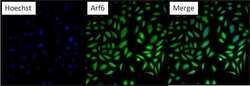
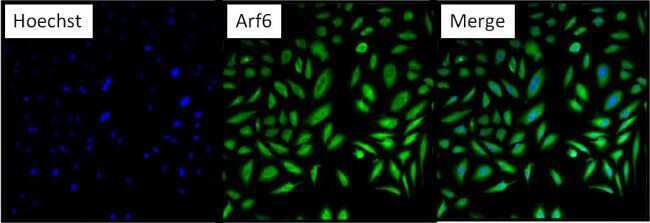
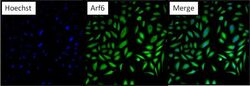
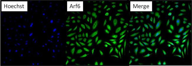
- Immunofluorescent analysis of Arf6 (green) in HeLa cells. Formalin fixed cells were permeabilized with 0.1% Triton X-100 in TBS for 10 minutes at room temperature and blocked with 5% normal goat serum (Product # 31873) for 15 minutes at room temperature. Cells were probed with an Arf6 polyclonal antibody (Product # PA1-093) at a dilution of 1:100 for at least 1 hour at room temperature, washed with PBS, and incubated with DyLight 488 goat anti-rabbit IgG secondary antibody (Product # 35552) at a dilution of 1:400 for 30 minutes at room temperature. Nuclei (blue) were stained with Hoechst 33342 dye (Product # 62249). Images were taken on a Thermo Scientific ArrayScan at 20X magnification.
- Submitted by
- Invitrogen Antibodies (provider)
- Main image

- Experimental details
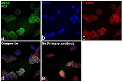
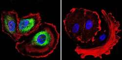
- Immunofluorescence analysis of ARF6 was performed using 70% confluent log phase PC-3 cells. The cells were fixed with 4% paraformaldehyde for 10 minutes, permeabilized with 0.1% Triton™ X-100 for 15 minutes, and blocked with 2% BSA for 45 minutes at room temperature. The cells were labeled with ARF6 Polyclonal Antibody (Product # PA1-093) at 1:100 in 0.1% BSA, incubated at 4 degree celsius overnight and then labeled with Donkey anti-Rabbit IgG (H+L) Highly Cross-Adsorbed Secondary Antibody, Alexa Fluor Plus 488 (Product # A32790), (1:2000), for 45 minutes at room temperature (Panel a: Green). Nuclei (Panel b:Blue) were stained with ProLong™ Diamond Antifade Mountant with DAPI (Product # P36962). F-actin (Panel c: Red) was stained with Rhodamine Phalloidin (Product # R415, 1:300). Panel d represents the merged image showing Cytoplasmic localization. Panel e represents control cells with no primary antibody to assess background. The images were captured at 40X magnification.

- Submitted by
- Invitrogen Antibodies (provider)
- Main image
- Experimental details
- Immunofluorescent analysis of Arf6 (green) in HeLa cells. Formalin fixed cells were permeabilized with 0.1% Triton X-100 in TBS for 10 minutes at room temperature and blocked with 5% normal goat serum (Product # 31873) for 15 minutes at room temperature. Cells were probed with an Arf6 polyclonal antibody (Product # PA1-093) at a dilution of 1:100 for at least 1 hour at room temperature, washed with PBS, and incubated with DyLight 488 goat anti-rabbit IgG secondary antibody (Product # 35552) at a dilution of 1:400 for 30 minutes at room temperature. Nuclei (blue) were stained with Hoechst 33342 dye (Product # 62249). Images were taken on a Thermo Scientific ArrayScan at 20X magnification.
- Submitted by
- Invitrogen Antibodies (provider)
- Main image
- Experimental details
- Immunofluorescent analysis of Arf6 in MCF-7 cells. Cells were grown on chamber slides and fixed with formaldehyde prior to staining. Cells were probed without (control) or with a Arf6 monoclonal antibody (Product # PA1-093) at a dilution of 1:20 overnight at 4 C, washed with PBS and incubated with a DyLight-488 conjugated secondary antibody (Product # 35552). Arf6 Polyclonal Antibody staining (green), F-Actin staining with Phalloidin (red) and nuclei with DAPI (blue) is shown. Images were taken at 60X magnification.

Supportive validation
- Submitted by
- Invitrogen Antibodies (provider)
- Main image
- Experimental details
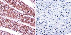
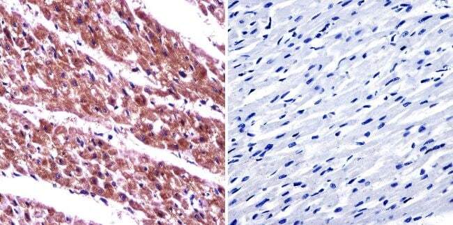
- Immunohistochemistry was performed on normal biopsies of deparaffinized human heart tissue. To expose target proteins, heat induced antigen retrieval was performed using 10mM sodium citrate (pH6.0) buffer, microwaved for 8-15 minutes. Following antigen retrieval tissues were blocked in 3% BSA-PBS for 30 minutes at room temperature. Tissues were then probed at a dilution of 1:20 with a Rabbit Polyclonal Antibody recognizing Arf6 (Product # PA1-093) or without primary antibody (negative control) overnight at 4°C in a humidified chamber. Tissues were washed extensively with PBST and endogenous peroxidase activity was quenched with a peroxidase suppressor. Detection was performed using a biotin-conjugated secondary antibody and SA-HRP, followed by colorimetric detection using DAB. Tissues were counterstained with hematoxylin and prepped for mounting.
Supportive validation
- Submitted by
- Invitrogen Antibodies (provider)
- Main image
- Experimental details
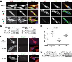
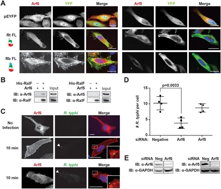
- Fig 6 Arf6 is recruited by R . typhi RalF and is required for infection. (A) Ectopically expressed RalF RtFL co-localizes with Arf6 but not Arf5. HeLa cells co-expressing EYFP, EYFP-RalF RtFL or EYFP-RalF RbFL and mRFP-Arf6 (left) or -Arf5 (right) were fixed with 4% para-formaldehyde. Nuclei were stained with DAPI (blue). (Scale bar: 10 mum). (B) RalF RtFL pull-down of Arf6. Lysates from HEK293T cells expressing mRFP-Arf5 or -Arf6 were incubated with HisPur Cobalt resin bound with rHis-RalF RtFL or resin alone. Bound proteins were eluted with imidazole and analyzed by protein immunoblot using antibodies as indicated. (C) Arf6 is recruited during R . typhi entry. HeLa cells expressing mRFP-Arf5 or -Arf6 (red) were infected with partially purified R . typhi (MOI ~100). Ten minutes post infection, cells were fixed and R . typhi detected with anti- R . typhi serum (green). DAPI (blue) is shown in the merged image. Boxed regions are enlarged to show detail. White arrowheads indicate R . typhi . (Scale bar: 5 mum). (D) Arf6 knockdown inhibits R . typhi infection. HeLa cells transfected with negative, Arf6, or Arf5 siRNA were infected with partially purified R . typhi (MOI ~100). At 2 hrs post infection, cells were fixed, plasma membrane stained with Alexa Fluor 594 wheat germ agglutinin, and R . typhi detected with rat anti- R . typhi serum and Alexa Fluor 488 anti-rat antibody. The number of R . typhi per host cell was counted for 100 host cells for three independent experiments.
- Submitted by
- Invitrogen Antibodies (provider)
- Main image
- Experimental details
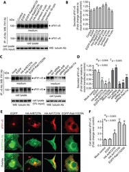
- Figure 3 Secretion of sFlt1 from endothelial cells requires functional Arf6, Arf1, and Rab11 GTPases. A-F : EC-sFlt1 cells were nucleofected with plasmid constructs expressing dominant-negative forms of Arf1, Arf6, Rab5, Rab6, Rab 8, syntaxin 4, syntaxin 6, and syntaxin 16. Similarly, in a separate study EC-sFlt1 cells were nucleofected with scramble oligonucleotide or siRNAs against Arf1 , Arf6 , and Rab11a . Media containing nucleofection complexes were replaced with fresh media 8 hrs post-nucleofection. At 36 hrs post-nucleofection, secreted and intracellular sFlt1 were immunoprecipitated from spent culture media and cell lysates using the anti-v5 Ab. ( A , C ) Representative blot shows relative expression of sFlt1-v5 in cell lysates and secreted in culture media. B , D : Band densities from experiments as in A, C were quantified by densitometric analysis. Secretion of sFlt1-v5 was calculated as the ratio of desitometric band intensity values of secreted to intracellular Flt1; this ratio was set at 1 for mock-transfected control EC-sFlt1 cells. The data are the means +- SEM from three independent experiments. E : EC-sFlt1 cells were transiently transfected with indicated dominant-negative plasmid constructs. After 24 hrs cells were fixed, permeabilized, and labeled with anti-v5 Ab to detect cell-associated sFlt1-v5. Representative images obtained by epifluorescence microscopy show localization of sFlt1-v5. F : Quantification of intracellular retention of sFlt1-v5 upon inhi

- Submitted by
- Invitrogen Antibodies (provider)
- Main image
- Experimental details
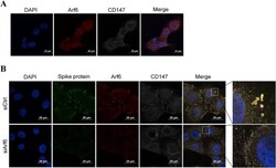
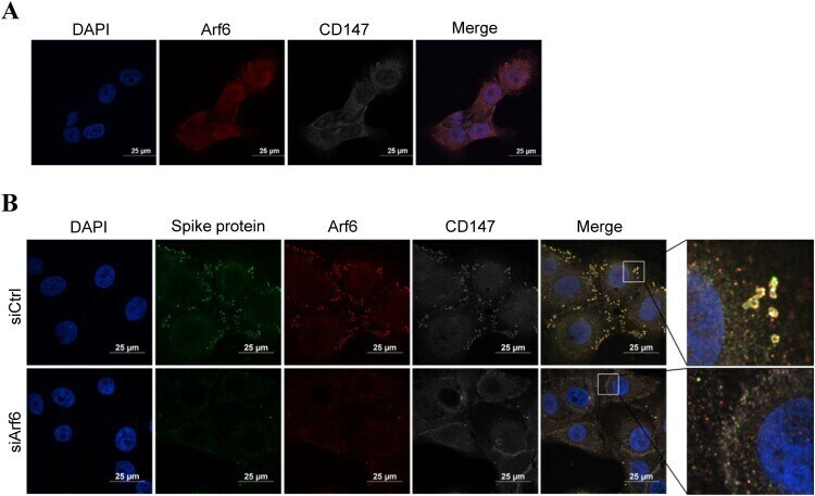
- Figure 4. The co-localization of targeted proteins was observed by immunofluorescence staining. (A) The co-localization between Arf6 (red) and CD147 (white) was analyzed in Vero E6 cells by immunofluorescence staining. Scale bars, 25 mum. (B) The co-localization of spike protein (green), Arf6 (red), and CD147 (white) was analyzed in Vero E6 cells and Arf6 knockdown cells by multicolour immunofluorescence staining. Scale bars, 25 mum.
- Submitted by
- Invitrogen Antibodies (provider)
- Main image
- Experimental details
- Figure 6. The levels of activated Arf6 in cortical tissue are elevated because of Iqsec2 KO. (A) Biochemical assays to measure the levels of activated Arf6 (G-LISA) undertaken in cortical tissues of animals across postnatal development between 2 and 9 mo of age show that (A) the levels of activated Arf6 in wild-type male mice (WT/black) (n = 4) are elevated in age-matched wild-type female mice (WT/grey) (n = 5). (B) Iqsec2 KO hemizygous males (KO/blue, n = 6) and heterozygous KO females (Het/pink; n = 9) both display increased levels of activated Arf6 compared with the sex-matched wild-type controls listed above. (C) The abundance of Arf6 protein measured by immunoblot was not significantly different between any genotype groups. The Iqsec2 KO animals with observed seizures are denoted by stars. Mean (+-SEM) data presented, where * indicates significance between WT female control and HET/KO, P < 0.05, 2-tailed, unpaired t test.
- Submitted by
- Invitrogen Antibodies (provider)
- Main image
- Experimental details
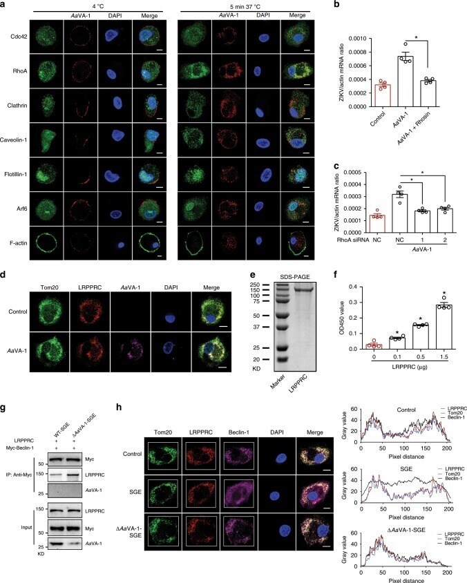
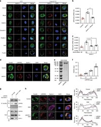
- Fig. 5 Aa VA-1 competes with Beclin-1 from the LRPPRC-mediated inhibition. a The localization of Aa VA-1 in human moMO cells. Purified Aa VA-1 (5 mug/mL) was incubated with human moMO cells at 4 degC or 37 degC. Both Aa VA-1 and specific markers of the endocytosis pathways were stained, and nuclei were stained with DAPI. Scale bars, 10 mum. b , c Suppression of RhoA by the antagonist Rhosin ( b ) and siRNA-mediated silencing ( c ) restored Aa VA-1-mediated ZIKV enhancement in THP-1 cells. Gene quantities were normalized against human actin ( NM_001101.4 ). d Colocalization of Aa VA-1 and LRPPRC on mitochondria in SGE-treated human moMO cells. Purified Aa VA-1 (biotin-labeled, 5 mug/mL) was incubated with human moMO for 6 h. The cells were stained with anti-LRPPRC and anti-Tom20, as well as streptavidin Alexa-633. The nuclei were stained with DAPI. Images were examined using a Zeiss LSM 780 meta confocal microscope in multi-track mode. Scale bars, 10 mum. e Purification of a recombinant LRPPRC protein in 293F cells. The protein was detected by staining with Coomassie blue in an SDS-PAGE gel. f Aa VA-1 directly binds LRPPRC. This interaction was detected by ELISA. g Aa VA-1 impaired the binding between LRPPRC and Beclin-1. SGE or Delta Aa VA-1-SGE was incubated with the lysates from Beclin-1 (Myc tag)-expressing cells to investigate protein interactions. The protein complex was pulled down with anti-Myc antibodies. h Treatment of SGE dissociated Beclin-1 from LRPPRC on mitochon