Explore
 Validate
 Learn
Learn Western blot
 Immunocytochemistry
Antibody data
- Antibody Data
- Antigen structure
- References [0]
- Comments [0]
- Validations
- Immunocytochemistry [4]
- Immunohistochemistry [1]
Submit
Validation data
Reference
Comment
Report error
- Product number
- 702787 - Provider product page

- Provider
- Invitrogen Antibodies
- Product name
- VAMP1 Recombinant Rabbit Monoclonal Antibody (24H9L10)
- Antibody type
- Monoclonal
- Antigen
- Synthetic peptide
- Description
- This antibody is predicted to react with Monkey, Mouse, Pig, Rabbit, Bovine. Recombinant rabbit monoclonal antibodies are produced using in vitro expression systems. The expression systems are developed by cloning in the specific antibody DNA sequences from immunoreactive rabbits. Then, individual clones are screened to select the best candidates for production. The advantages of using recombinant rabbit monoclonal antibodies include: better specificity and sensitivity, lot-to-lot consistency, animal origin-free formulations, and broader immunoreactivity to diverse targets due to larger rabbit immune repertoire.
- Reactivity
- Human, Mouse, Rat
- Host
- Rabbit
- Isotype
- IgG
- Antibody clone number
- 24H9L10
- Vial size
- 100 μg
- Concentration
- 0.5 mg/mL
- Storage
- Store at 4°C short term. For long term storage, store at -20°C, avoiding freeze/thaw cycles.
No comments: Submit comment
Supportive validation
- Submitted by
- Invitrogen Antibodies (provider)
- Main image
- Experimental details
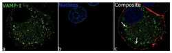
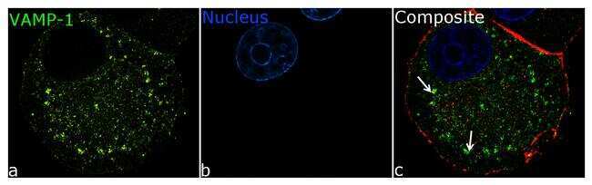
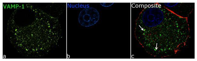
- For immunofluorescence analysis, PC12 cells were fixed and permeabilized for detection of endogenous VAMP1 using Anti- VAMP1 Recombinant Rabbit Monoclonal Antibody (Product # 702787, 5 µg/mL) and labeled with Goat anti-Rabbit IgG (H+L) Superclonal™ Secondary Antibody, Alexa Fluor® 488 conjugate (Product # A27034, 1:2000). Panel a) shows representative cells that were stained for detection and localization of VAMP1 protein (green), Panel b) is stained for nuclei (blue) using SlowFade® Gold Antifade Mountant with DAPI (Product # S36938). anel c) is a composite image of Panels a and b clearly demonstrating localisation of VAMP-1 in cytoplamic vesicles. The images were captured at 100X magnification.
- Submitted by
- Invitrogen Antibodies (provider)
- Main image
- Experimental details
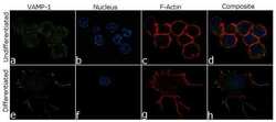
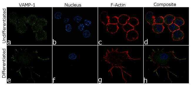
- For immunofluorescence analysis, PC12 cells were fixed and permeabilized for detection of endogenous VAMP1 using Anti-VAMP1 Recombinant Rabbit Monoclonal Antibody (Product # 702787, 5 µg/mL) and labeled with Goat anti-Rabbit IgG (H+L) Superclonal™ Secondary Antibody, Alexa Fluor® 488 conjugate (Product # A27034, 1:2000). Nuclei (blue) is stained using SlowFade® Gold Antifade Mountant with DAPI (Product # S36938) and cytoskeletal F-actin (red) staining using Rhodamine Phalloidin (Product # R415, 1:300) Panel a-d) shows representative undifferentiated cells that were stained for detection and localization of VAMP1 protein (green) with no signal. Panel e-h) clearly demonstrate enhanced signal of VMAP1 in cell body and neuronal processes of NGF (200 ng/mL 5days) induced differentiated PC12 cells. The images were captured at 60X magnification.
- Submitted by
- Invitrogen Antibodies (provider)
- Main image

- Experimental details
- For immunofluorescence analysis, PC12 cells were fixed and permeabilized for detection of endogenous VAMP1 using Anti- VAMP1 Recombinant Rabbit Monoclonal Antibody (Product # 702787, 5 µg/mL) and labeled with Goat anti-Rabbit IgG (Heavy Chain) Superclonal™ Secondary Antibody, Alexa Fluor® 488 conjugate (Product # A27034, 1:2000). Panel a) shows representative cells that were stained for detection and localization of VAMP1 protein (green), Panel b) is stained for nuclei (blue) using SlowFade® Gold Antifade Mountant with DAPI (Product # S36938). anel c) is a composite image of Panels a and b clearly demonstrating localisation of VAMP-1 in cytoplamic vesicles. The images were captured at 100X magnification.
- Submitted by
- Invitrogen Antibodies (provider)
- Main image
- Experimental details
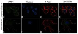
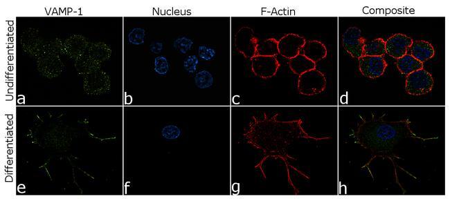
- For immunofluorescence analysis, PC12 cells were fixed and permeabilized for detection of endogenous VAMP1 using Anti-VAMP1 Recombinant Rabbit Monoclonal Antibody (Product # 702787, 5 µg/mL) and labeled with Goat anti-Rabbit IgG (Heavy Chain) Superclonal™ Secondary Antibody, Alexa Fluor® 488 conjugate (Product # A27034, 1:2000). Nuclei (blue) is stained using SlowFade® Gold Antifade Mountant with DAPI (Product # S36938) and cytoskeletal F-actin (red) staining using Rhodamine Phalloidin (Product # R415, 1:300) Panel a-d) shows representative undifferentiated cells that were stained for detection and localization of VAMP1 protein (green) with no signal. Panel e-h) clearly demonstrate enhanced signal of VMAP1 in cell body and neuronal processes of NGF (200 ng/mL 5days) induced differentiated PC12 cells. The images were captured at 60X magnification.
Supportive validation
- Submitted by
- Invitrogen Antibodies (provider)
- Main image

- Experimental details
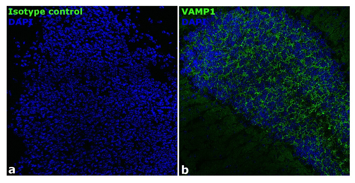
- Immunofluorescence analysis of mouse brain tissue: Frozen sections were fixed with 4% PFA for 20 min, permeabilized using 0.1% Triton-X 100 for 10 mins and blocked for 1 hour with 2% BSA. Transverse sections of mouse brain were incubated with Anti-VAMP1 Recombinant Rabbit Polyclonal Antibody (Product # 702787, 1:250 dilution) overnight at 4°C, followed by Goat anti-Rabbit IgG (Heavy Chain) Superclonal™ Secondary Antibody, Alexa Fluor® 488 conjugate (Product # A27034, 1:2000, 45 mins). Nuclei (blue) were stained using SlowFade® Gold Antifade Mountant with DAPI (Product # S36938), Panel a) represents staining with the matched isotype control. Panel b) shows a representative hippocampal region stained for VAMP1 (green). The images were captured at 20X magnification.