 Explore
 Validate
 Learn
Learn Western blot
Antibody data
- Antibody Data
- Antigen structure
- References [0]
- Comments [0]
- Validations
- Western blot [8]
- Immunocytochemistry [1]
Submit
Validation data
Reference
Comment
Report error
- Product number
- PA5-85552 - Provider product page

- Provider
- Invitrogen Antibodies
- Product name
- PROX1 Polyclonal Antibody
- Antibody type
- Polyclonal
- Antigen
- Recombinant full-length protein
- Description
- Keep as concentrated solution. Predicted reactivity: Mouse (97%), Rat (97%), Pig (97%), Chicken (89%), Rhesus Monkey (100%), Bovine (97%). Positive Control: HepG2 nuclear extract, mouse hippocampus, mouse eye, fetal mouse forebrain. Store product as a concentrated solution. Centrifuge briefly prior to opening the vial.
- Reactivity
- Human, Mouse, Rat
- Host
- Rabbit
- Isotype
- IgG
- Vial size
- 100 µL
- Concentration
- 1 mg/mL
- Storage
- Store at 4°C short term. For long term storage, store at -20°C, avoiding freeze/thaw cycles.
No comments: Submit comment
Supportive validation
- Submitted by
- Invitrogen Antibodies (provider)
- Main image
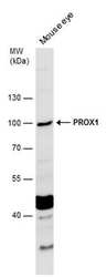
- Experimental details
- Western blot analysis of PROX1 in mouse tissue extract using PROX1 polyclonal antibody (Product # PA5-85552) using 50 µg of sample at a dilution of 1:1000. Sample was then incubated with HRP-conjugated anti-rabbit IgG secondary antibody. Prior to incubation with primary antibody, the sample was separated on 7.5% SDS-PAGE.
- Submitted by
- Invitrogen Antibodies (provider)
- Main image
- Experimental details
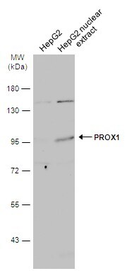
- Western blot analysis of PROX1 in HepG2 whole cell extract and conditioned medium using PROX1 polyclonal antibody (Product # PA5-85552) using 30 µg of sample at a dilution of 1:500. Sample was then incubated with HRP-conjugated anti-rabbit IgG secondary antibody. Prior to incubation with primary antibody, the sample was separated on 7.5% SDS-PAGE.
- Submitted by
- Invitrogen Antibodies (provider)
- Main image
- Experimental details
- Western blot analysis of PROX1 in A) 50 µg fetal mouse forebrain lysate using PROX1 polyclonal antibody (Product # PA5-85552) using 50 µg of sample at a dilution of 1:1000. Prior to incubation with primary antibody, the sample was separated on 7.5% SDS-PAGE.

- Submitted by
- Invitrogen Antibodies (provider)
- Main image
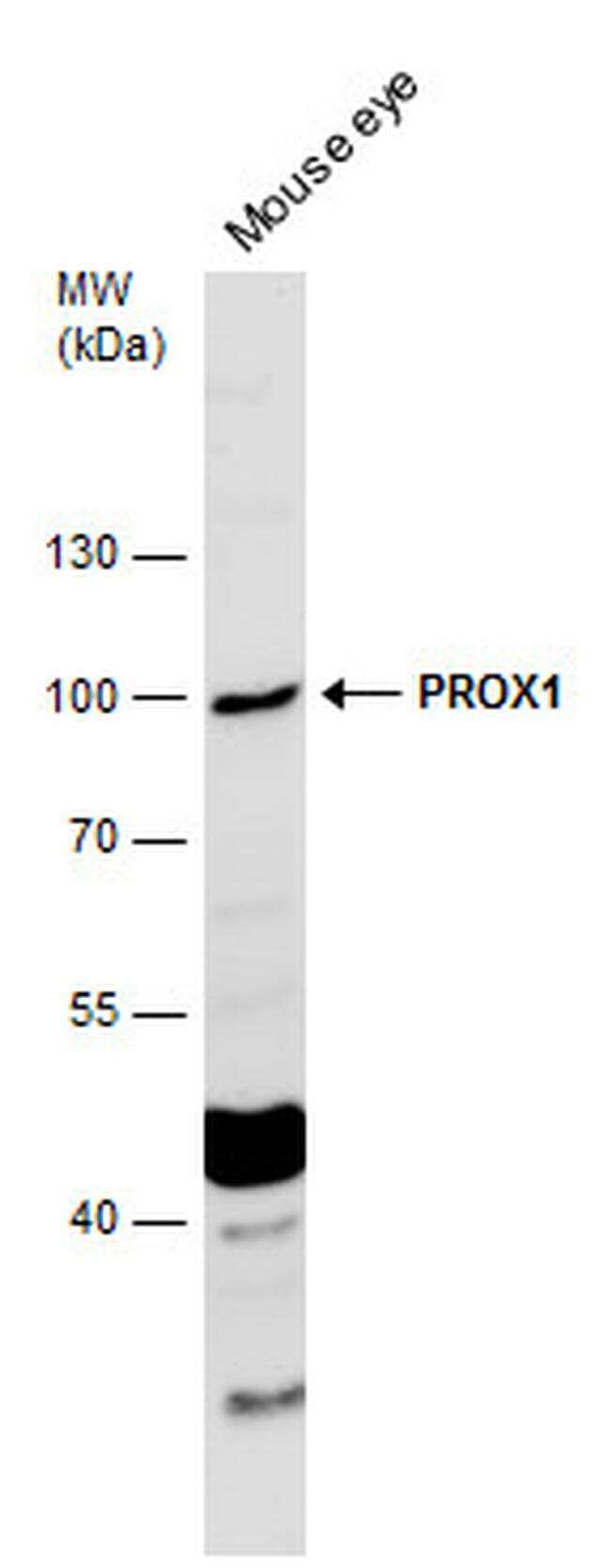
- Experimental details
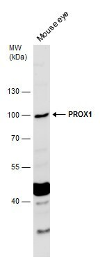
- Western blot analysis of PROX1 in mouse tissue extract using PROX1 polyclonal antibody (Product # PA5-85552) using 50 µg of sample at a dilution of 1:500. Prior to incubation with primary antibody, the sample was separated on 7.5% SDS-PAGE.
- Submitted by
- Invitrogen Antibodies (provider)
- Main image
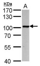
- Experimental details
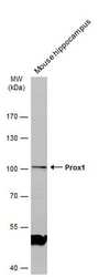
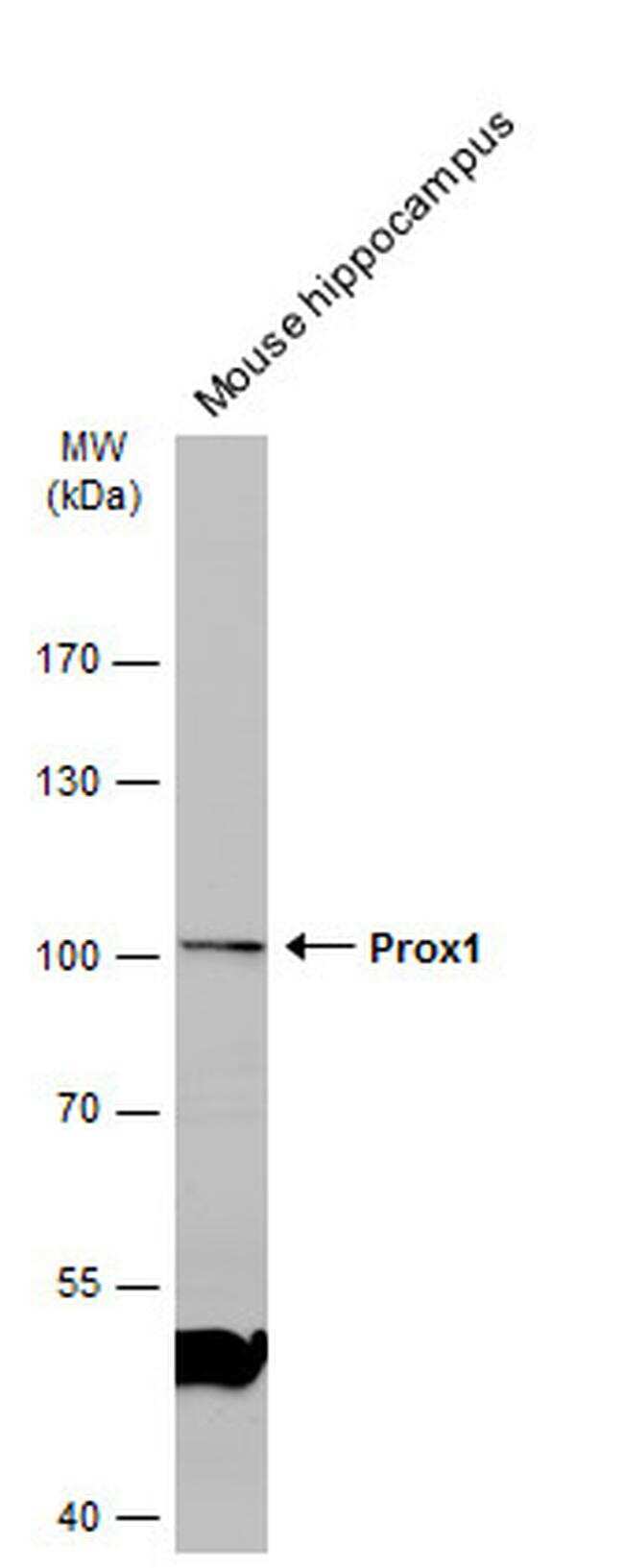
- Western Blot using PROX1 Polyclonal Antibody (Product # PA5-85552). Mouse tissue extract (50 µg) was separated by 7.5% SDS-PAGE, and the membrane was blotted with PROX1 Polyclonal Antibody (Product # PA5-85552) diluted at 1:1,000. The HRP-conjugated anti-rabbit IgG antibody was used to detect the primary antibody.

- Submitted by
- Invitrogen Antibodies (provider)
- Main image
- Experimental details
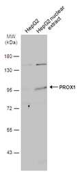
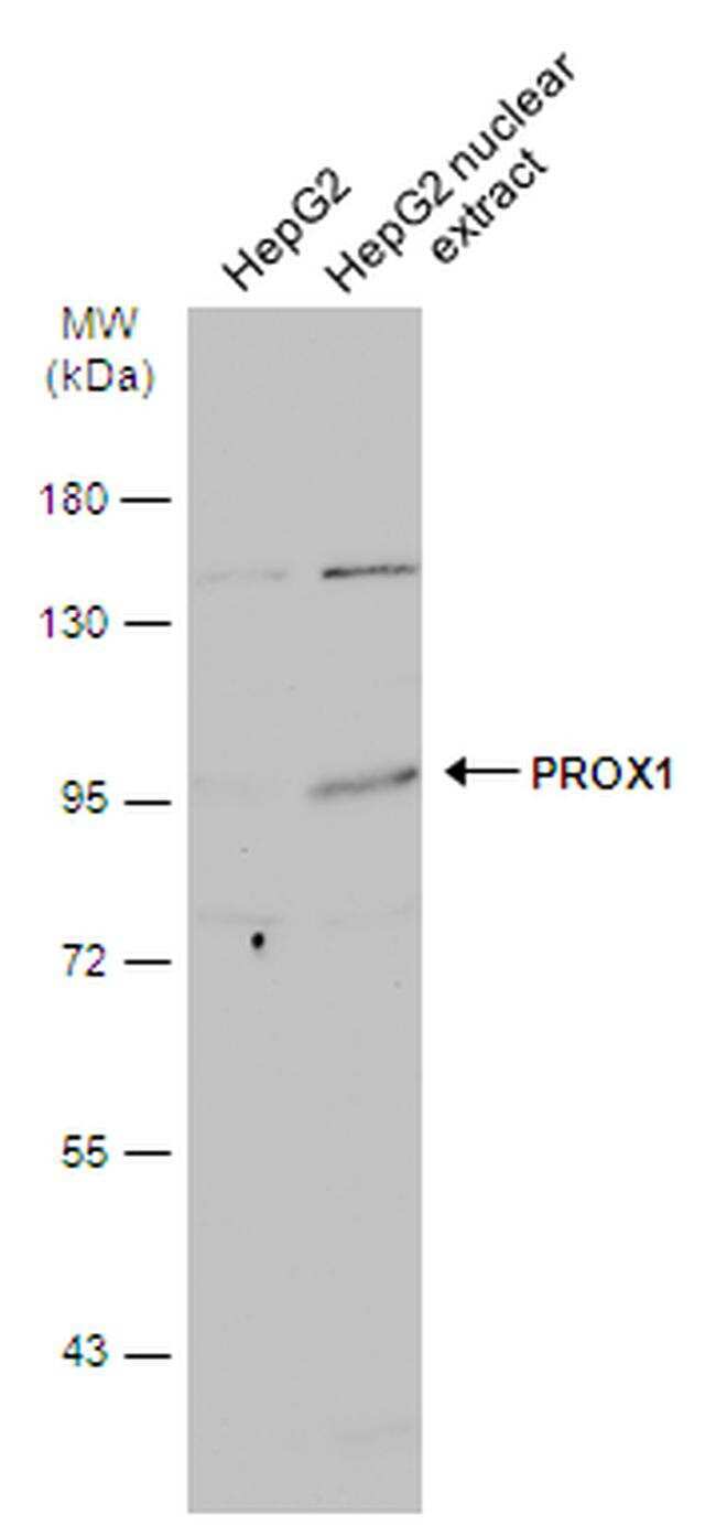
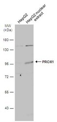
- Western Blot using PROX1 Polyclonal Antibody (Product # PA5-85552). HepG2 whole cell and nuclear extracts (30 µg) were separated by 7.5% SDS-PAGE, and the membrane was blotted with PROX1 Polyclonal Antibody (Product # PA5-85552) diluted at 1:500. The HRP-conjugated anti-rabbit IgG antibody was used to detect the primary antibody.
- Submitted by
- Invitrogen Antibodies (provider)
- Main image
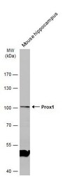
- Experimental details
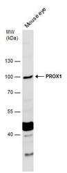
- Western Blot using PROX1 Polyclonal Antibody (Product # PA5-85552). Mouse tissue extract (50 µg) was separated by 7.5% SDS-PAGE, and the membrane was blotted with PROX1 Polyclonal Antibody (Product # PA5-85552) diluted at 1:500.
- Submitted by
- Invitrogen Antibodies (provider)
- Main image

- Experimental details
- PROX1 Polyclonal Antibody detects PROX1 protein by western blot analysis. A. 50 µg fetal mouse forebrain lysate/extract.7.5 % SDS-PAGE. PROX1 Polyclonal Antibody (Product # PA5-85552) dilution: 1:1,000.

Supportive validation
- Submitted by
- Invitrogen Antibodies (provider)
- Main image
- Experimental details
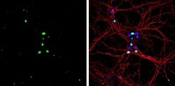
- PROX1 Polyclonal Antibody detects PROX1 protein by immunofluorescent analysis. Sample: DIV10 rat E18 primary hippocampal neurons were fixed in 4% paraformaldehyde at RT for 15 min. Green: PROX1 protein stained by PROX1 Polyclonal Antibody (Product # PA5-85552) diluted at 1:500. Red: beta Tubulin 3/ Tuj1, stained by beta Tubulin 3/ Tuj1 antibody [GT1338] diluted at 1:500. Blue: Fluoroshield with DAPI .
