 Explore
 Validate
 Learn
Learn ELISA
ELISA Immunohistochemistry
Antibody data
- Antibody Data
- Antigen structure
- References [2]
- Comments [0]
- Validations
- Immunohistochemistry [3]
- Flow cytometry [1]
Submit
Validation data
Reference
Comment
Report error
- Product number
- NBP1-30045 - Provider product page

- Provider
- Novus Biologicals
- Proper citation
- Novus Cat#NBP1-30045, RRID:AB_1968604
- Product name
- Mouse Monoclonal Prox1 Antibody
- Antibody type
- Monoclonal
- Description
- Protein G purified.
- Reactivity
- Human, Mouse, Rat, Chicken/Avian
- Host
- Mouse
- Antigen sequence
The homeo prospero domain of Prox1- Isotype
- IgG
- Vial size
- 0.1 ml
- Concentration
- 1.0 mg/ml
- Storage
- Store at 4C short term. Aliquot and store at -20C long term. Avoid freeze-thaw cycles.
Submitted references Human organotypic lymphatic vessel model elucidates microenvironment-dependent signaling and barrier function.
VEGF-C induces lymphangiogenesis and angiogenesis in the rat mesentery culture model.
Gong MM, Lugo-Cintron KM, White BR, Kerr SC, Harari PM, Beebe DJ
Biomaterials 2019 Sep;214:119225
Biomaterials 2019 Sep;214:119225
VEGF-C induces lymphangiogenesis and angiogenesis in the rat mesentery culture model.
Sweat RS, Sloas DC, Murfee WL
Microcirculation (New York, N.Y. : 1994) 2014 Aug;21(6):532-40
Microcirculation (New York, N.Y. : 1994) 2014 Aug;21(6):532-40
No comments: Submit comment
Supportive validation
- Submitted by
- Novus Biologicals (provider)
- Main image

- Experimental details
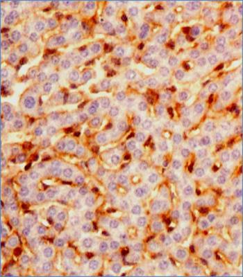
- Immunohistochemistry-Paraffin: Prox1 Antibody (5G10) [NBP1-30045] - IHC analysis of a formalin fixed paraffin embedded tissue section of Mouse liver using Lot UD1009o of PROX1 antibody clone 5G10 at 1:100 dilution with HRP-DAB detection and hematoxylin counterstaining. The antibody generated nice nuclear-cytoplasmic staining in sinusoidal endothelial cells as well as in Kupffer cells.
- Submitted by
- Novus Biologicals (provider)
- Main image
- Experimental details
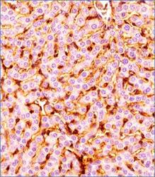
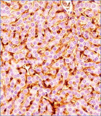
- Immunohistochemistry-Paraffin: Prox1 Antibody (5G10) [NBP1-30045] - IHC analysis of a formalin fixed paraffin embedded tissue section of Mouse liver using Lot A of PROX1 antibody clone 5G10 at 1:100 dilution with HRP-DAB detection and hematoxylin counterstaining. The antibody generated nice nuclear-cytoplasmic staining in sinusoidal endothelial cells as well as in Kupffer cells.
- Submitted by
- Novus Biologicals (provider)
- Main image
- Experimental details
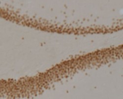
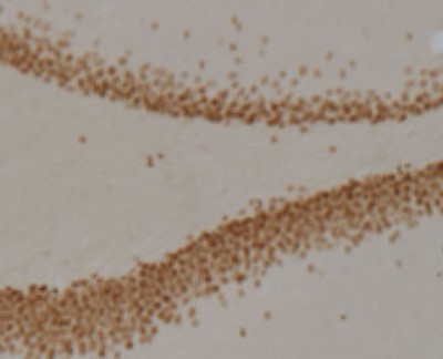
- Immunohistochemistry: Prox1 Antibody (5G10) [NBP1-30045] - Rat dentate gyrus showing specific immunolabeling of the prox1 protein. Photo courtesy of Justin Kievits and Teresa Milner, Weill Cornell Medical College.
Supportive validation
- Submitted by
- Novus Biologicals (provider)
- Main image
- Experimental details
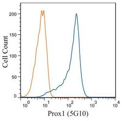
- Flow Cytometry: Prox1 Antibody (5G10) [NBP1-30045] - HepG2 cells were stained with Prox1 (5G10) NBP1-30045 (blue) and a matched isotype control NBP2-27287 (orange). Cells were fixed with 4% PFA and then permeablized with 0.1% saponin. Cells were incubated in an antibody dilution of 1 ug/mL for 30 minutes at room temperature, followed by Dylight488-conjugated anti-mouse secondary antibody.
