Explore
 Validate
 Learn
Learn Western blot
Antibody data
- Antibody Data
- Antigen structure
- References [5]
- Comments [0]
- Validations
- Western blot [1]
- Immunocytochemistry [1]
Submit
Validation data
Reference
Comment
Report error
- Product number
- MAB3059 - Provider product page

- Provider
- R&D Systems
- Product name
- Mouse S100A8 Antibody
- Antibody type
- Monoclonal
- Description
- Protein A or G purified from hybridoma culture supernatant. Detects mouse S100A8 in direct ELISAs and Western blots. In direct ELISAs and Western blots, 40% cross-reactivity with recombinant human (rh) S100A8 is observed and no cross-reactivity with recombinant mouse (rm) S100A4, rmS100A9, rmS100A10, rmS100A11, rhS100B, or rhS100P is observed.
- Reactivity
- Mouse
- Host
- Rat
- Conjugate
- Unconjugated
- Antigen sequence
P27005- Isotype
- IgG
- Antibody clone number
- 335806
- Vial size
- 100 ug
- Concentration
- LYOPH
- Storage
- Use a manual defrost freezer and avoid repeated freeze-thaw cycles. 12 months from date of receipt, -20 to -70 °C as supplied. 1 month, 2 to 8 °C under sterile conditions after reconstitution. 6 months, -20 to -70 °C under sterile conditions after reconstitution.
Submitted references A Critical Role of Zinc Importer AdcABC in Group A Streptococcus-Host Interactions During Infection and Its Implications for Vaccine Development.
The calcium-binding protein complex S100A8/A9 has a crucial role in controlling macrophage-mediated renal repair following ischemia/reperfusion.
Visualized macrophage dynamics and significance of S100A8 in obese fat.
The acute neutrophil response mediated by S100 alarmins during vaginal Candida infections is independent of the Th17-pathway.
S100A8/A9 is not involved in host defense against murine urinary tract infection.
Makthal N, Nguyen K, Do H, Gavagan M, Chandrangsu P, Helmann JD, Olsen RJ, Kumaraswami M
EBioMedicine 2017 Jul;21:131-141
EBioMedicine 2017 Jul;21:131-141
The calcium-binding protein complex S100A8/A9 has a crucial role in controlling macrophage-mediated renal repair following ischemia/reperfusion.
Dessing MC, Tammaro A, Pulskens WP, Teske GJ, Butter LM, Claessen N, van Eijk M, van der Poll T, Vogl T, Roth J, Florquin S, Leemans JC
Kidney international 2015 Jan;87(1):85-94
Kidney international 2015 Jan;87(1):85-94
Visualized macrophage dynamics and significance of S100A8 in obese fat.
Sekimoto R, Fukuda S, Maeda N, Tsushima Y, Matsuda K, Mori T, Nakatsuji H, Nishizawa H, Kishida K, Kikuta J, Maijima Y, Funahashi T, Ishii M, Shimomura I
Proceedings of the National Academy of Sciences of the United States of America 2015 Apr 21;112(16):E2058-66
Proceedings of the National Academy of Sciences of the United States of America 2015 Apr 21;112(16):E2058-66
The acute neutrophil response mediated by S100 alarmins during vaginal Candida infections is independent of the Th17-pathway.
Yano J, Kolls JK, Happel KI, Wormley F, Wozniak KL, Fidel PL Jr
PloS one 2012;7(9):e46311
PloS one 2012;7(9):e46311
S100A8/A9 is not involved in host defense against murine urinary tract infection.
Dessing MC, Butter LM, Teske GJ, Claessen N, van der Loos CM, Vogl T, Roth J, van der Poll T, Florquin S, Leemans JC
PloS one 2010 Oct 14;5(10):e13394
PloS one 2010 Oct 14;5(10):e13394
No comments: Submit comment
Supportive validation
- Submitted by
- R&D Systems (provider)
- Main image

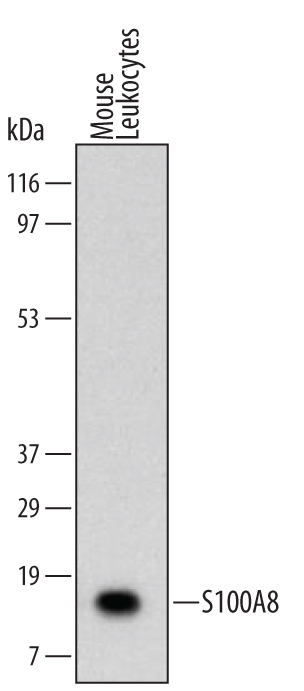
- Experimental details
- Detection of S100A8 Rat by Western Blot. Western blot shows lysates of mouse leukocyte. PVDF membrane was probed with 2 µg/mL of Rat Anti-Mouse S100A8 Monoclonal Antibody (Catalog # MAB3059) followed by HRP-conjugated Anti-Rat IgG Secondary Antibody (Catalog # HAF005). A specific band was detected for S100A8 at approximately 10 kDa (as indicated). This experiment was conducted under reducing conditions and using Immunoblot Buffer Group 1.
Supportive validation
- Submitted by
- R&D Systems (provider)
- Main image

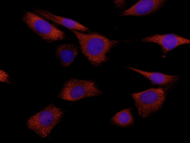
- Experimental details
- S100A8 in NIH-3T3 Mouse Cell Line. S100A8 was detected in immersion fixed NIH-3T3 mouse embryonic fibroblast cell line using 10 µg/mL Rat Anti-Mouse S100A8 Monoclonal Antibody (Catalog # MAB3059) for 3 hours at room temperature. Cells were stained (red) and counterstained with DAPI (blue). View our protocol for Fluorescent ICC Staining of Cells on Coverslips.