Explore
 Validate
 Learn
Learn Western blot
 Other assay
Antibody data
- Antibody Data
- Antigen structure
- References [35]
- Comments [0]
- Validations
- Other assay [27]
Submit
Validation data
Reference
Comment
Report error
- Product number
- 32-6300 - Provider product page

- Provider
- Invitrogen Antibodies
- Product name
- Desmoglein 3 Monoclonal Antibody (5G11)
- Antibody type
- Monoclonal
- Antigen
- Other
- Reactivity
- Human
- Host
- Mouse
- Isotype
- IgG
- Antibody clone number
- 5G11
- Vial size
- 100 μg
- Concentration
- 0.5 mg/mL
- Storage
- -20°C
Submitted references The Actin-Binding Protein α-Adducin Modulates Desmosomal Turnover and Plasticity.
Clustering of desmosomal cadherins by desmoplakin is essential for cell-cell adhesion.
In vitro Characteristics of Heterogeneous Equine Hoof Progenitor Cell Isolates.
The cell-cell junctions of mammalian testes: II. The lamellar smooth muscle monolayer cells of the peritubular wall are laterally connected by vertical adherens junctions-a novel architectonic cell-cell junction system.
Generation of an anti-desmoglein 3 antibody without pathogenic activity of pemphigus vulgaris for therapeutic application to squamous cell carcinoma.
Pemphigus Vulgaris Persistently Localized to the Nose with Local and Systemic Response to Topical Steroids.
Stat3 regulates desmoglein 3 transcription in epithelial keratinocytes.
A non-canonical role for desmoglein-2 in endothelial cells: implications for neoangiogenesis.
MAPKAP kinase 2 (MK2)-dependent and -independent models of blister formation in pemphigus vulgaris.
DSG3 facilitates cancer cell growth and invasion through the DSG3-plakoglobin-TCF/LEF-Myc/cyclin D1/MMP signaling pathway.
Desmoglein 2 is less important than desmoglein 3 for keratinocyte cohesion.
Pathological changes of the anatomical structure and markers of the limbal stem cell niche due to inflammation.
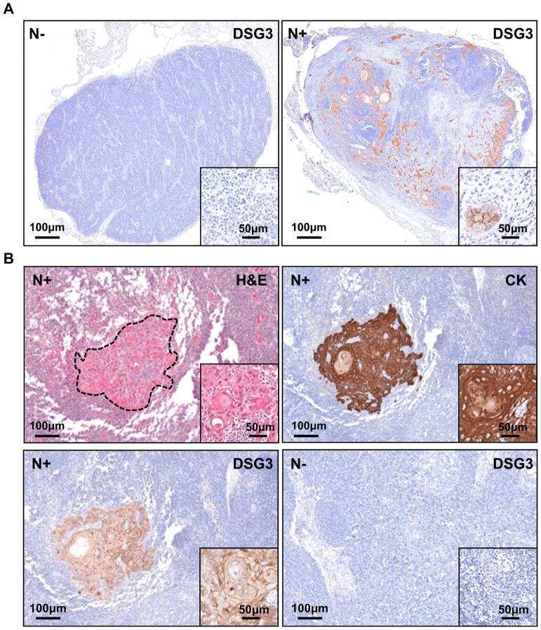
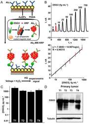
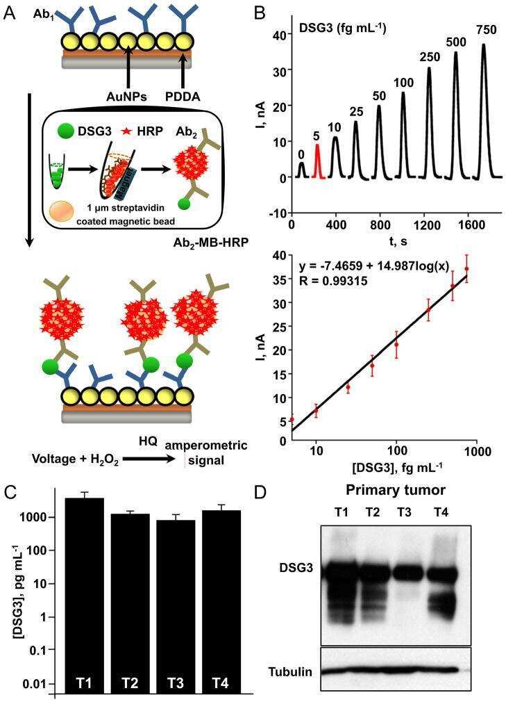
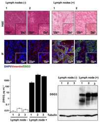
DSG3 as a biomarker for the ultrasensitive detection of occult lymph node metastasis in oral cancer using nanostructured immunoarrays.
Assessment of desmosomal components (desmoglein 1-3, plakoglobin) in cardia mucosa in relation to gastroesophageal reflux disease and Helicobacter pylori infection.
Gastro-oesophageal reflux disease is associated with up-regulation of desmosomal components in oesophageal mucosa.
Hailey-Hailey disease and tight junctions: Claudins 1 and 4 are regulated by ATP2C1 gene encoding Ca(2+) /Mn(2+) ATPase SPCA1 in cultured keratinocytes.
p38 MAPK activation is downstream of the loss of intercellular adhesion in pemphigus vulgaris.
Desmoglein 2-mediated adhesion is required for intestinal epithelial barrier integrity.
p38MAPK signaling and desmoglein-3 internalization are linked events in pemphigus acantholysis.
DNMT1 maintains progenitor function in self-renewing somatic tissue.
Autoimmunity to desmocollin 3 in pemphigus vulgaris.
Human eccrine sweat gland cells can reconstitute a stratified epidermis.
Infant limbus: an immunohistological study.
Disruption of desmosome assembly by monovalent human pemphigus vulgaris monoclonal antibodies.
P120 catenin is associated with desmogleins when desmosomes are assembled in high-Ca2+ medium but not when disassembled in low-Ca2+ medium in DJM-1 cells.
Pemphigus vulgaris immunoglobulin G can recognize a 130 000 MW antigen other than desmoglein 3 on peripheral blood mononuclear cell surface.
DSG3 is overexpressed in head neck cancer and is a potential molecular target for inhibition of oncogenesis.
Characterization of extracellular matrix components in the limbal epithelial stem cell compartment.
New skin-equivalent model from de-epithelialized amnion membrane.
Desmoglein endocytosis and desmosome disassembly are coordinated responses to pemphigus autoantibodies.
Genetic and functional characterization of human pemphigus vulgaris monoclonal autoantibodies isolated by phage display.
Molecular classification of head and neck squamous cell carcinomas using patterns of gene expression.
Molecular classification of head and neck squamous cell carcinomas using patterns of gene expression.
Expression of desmosomal proteins in squamous cell carcinomas of the skin.
Expression of desmosomal proteins in squamous cell carcinomas of the skin.
Hiermaier M, Kliewe F, Schinner C, Stüdle C, Maly IP, Wanuske MT, Rötzer V, Endlich N, Vielmuth F, Waschke J, Spindler V
The Journal of investigative dermatology 2021 May;141(5):1219-1229.e11
The Journal of investigative dermatology 2021 May;141(5):1219-1229.e11
Clustering of desmosomal cadherins by desmoplakin is essential for cell-cell adhesion.
Wanuske MT, Brantschen D, Schinner C, Stüdle C, Walter E, Hiermaier M, Vielmuth F, Waschke J, Spindler V
Acta physiologica (Oxford, England) 2021 Apr;231(4):e13609
Acta physiologica (Oxford, England) 2021 Apr;231(4):e13609
In vitro Characteristics of Heterogeneous Equine Hoof Progenitor Cell Isolates.
Yang Q, Pinto VMR, Duan W, Paxton EE, Dessauer JH, Ryan W, Lopez MJ
Frontiers in bioengineering and biotechnology 2019;7:155
Frontiers in bioengineering and biotechnology 2019;7:155
The cell-cell junctions of mammalian testes: II. The lamellar smooth muscle monolayer cells of the peritubular wall are laterally connected by vertical adherens junctions-a novel architectonic cell-cell junction system.
Domke LM, Franke WW
Cell and tissue research 2019 Feb;375(2):451-482
Cell and tissue research 2019 Feb;375(2):451-482
Generation of an anti-desmoglein 3 antibody without pathogenic activity of pemphigus vulgaris for therapeutic application to squamous cell carcinoma.
Funahashi SI, Kawai S, Fujii E, Taniguchi K, Nakano K, Ishikawa S, Aburatani H, Suzuki M
Journal of biochemistry 2018 Dec 1;164(6):471-481
Journal of biochemistry 2018 Dec 1;164(6):471-481
Pemphigus Vulgaris Persistently Localized to the Nose with Local and Systemic Response to Topical Steroids.
Zhang C, Goldscheider I, Ruzicka T, Sárdy M
Acta dermato-venereologica 2017 Oct 2;97(9):1136-1137
Acta dermato-venereologica 2017 Oct 2;97(9):1136-1137
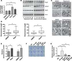
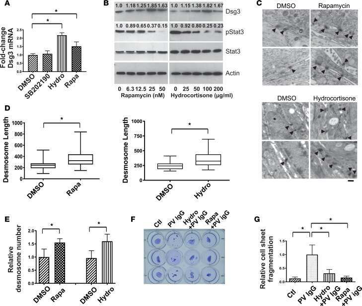
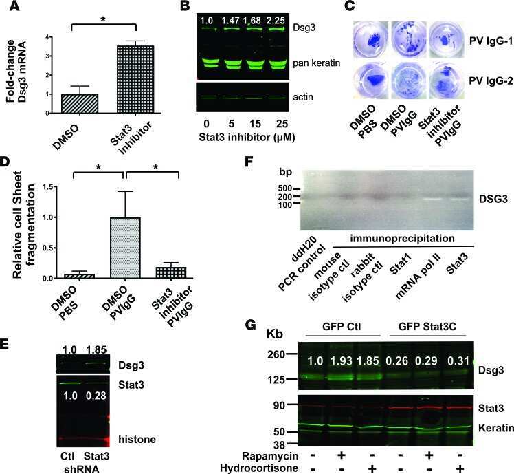
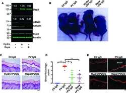
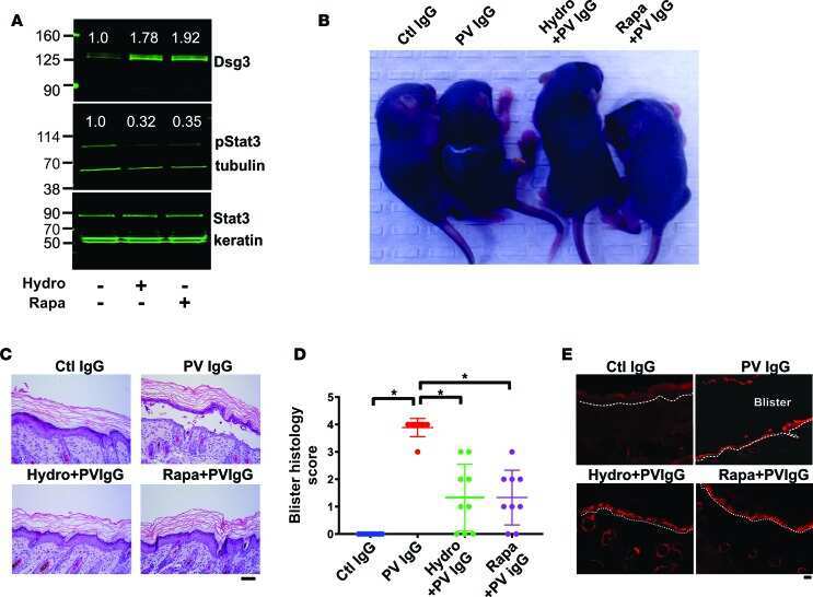
Stat3 regulates desmoglein 3 transcription in epithelial keratinocytes.
Mao X, Cho MJT, Ellebrecht CT, Mukherjee EM, Payne AS
JCI insight 2017 May 4;2(9)
JCI insight 2017 May 4;2(9)
A non-canonical role for desmoglein-2 in endothelial cells: implications for neoangiogenesis.
Ebert LM, Tan LY, Johan MZ, Min KK, Cockshell MP, Parham KA, Betterman KL, Szeto P, Boyle S, Silva L, Peng A, Zhang Y, Ruszkiewicz A, Zannettino AC, Gronthos S, Koblar S, Harvey NL, Lopez AF, Shackleton M, Bonder CS
Angiogenesis 2016 Oct;19(4):463-86
Angiogenesis 2016 Oct;19(4):463-86
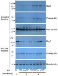
MAPKAP kinase 2 (MK2)-dependent and -independent models of blister formation in pemphigus vulgaris.
Mao X, Li H, Sano Y, Gaestel M, Mo Park J, Payne AS
The Journal of investigative dermatology 2014 Jan;134(1):68-76
The Journal of investigative dermatology 2014 Jan;134(1):68-76
DSG3 facilitates cancer cell growth and invasion through the DSG3-plakoglobin-TCF/LEF-Myc/cyclin D1/MMP signaling pathway.
Chen YJ, Lee LY, Chao YK, Chang JT, Lu YC, Li HF, Chiu CC, Li YC, Li YL, Chiou JF, Cheng AJ
PloS one 2013;8(5):e64088
PloS one 2013;8(5):e64088
Desmoglein 2 is less important than desmoglein 3 for keratinocyte cohesion.
Hartlieb E, Kempf B, Partilla M, Vigh B, Spindler V, Waschke J
PloS one 2013;8(1):e53739
PloS one 2013;8(1):e53739
Pathological changes of the anatomical structure and markers of the limbal stem cell niche due to inflammation.
Nubile M, Curcio C, Dua HS, Calienno R, Lanzini M, Iezzi M, Mastropasqua R, Agnifili L, Mastropasqua L
Molecular vision 2013;19:516-25
Molecular vision 2013;19:516-25
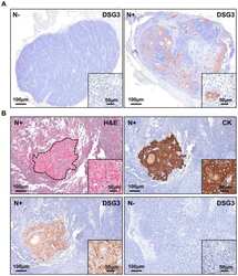
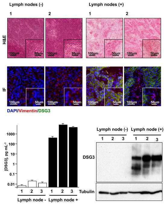
DSG3 as a biomarker for the ultrasensitive detection of occult lymph node metastasis in oral cancer using nanostructured immunoarrays.
Patel V, Martin D, Malhotra R, Marsh CA, Doçi CL, Veenstra TD, Nathan CA, Sinha UK, Singh B, Molinolo AA, Rusling JF, Gutkind JS
Oral oncology 2013 Feb;49(2):93-101
Oral oncology 2013 Feb;49(2):93-101
Assessment of desmosomal components (desmoglein 1-3, plakoglobin) in cardia mucosa in relation to gastroesophageal reflux disease and Helicobacter pylori infection.
Wex T, Kuester D, Mönkemüller K, Stahr A, Fry LC, Kandulski A, Kropf S, Roessner A, Malfertheiner P
Human pathology 2012 Oct;43(10):1745-54
Human pathology 2012 Oct;43(10):1745-54
Gastro-oesophageal reflux disease is associated with up-regulation of desmosomal components in oesophageal mucosa.
Wex T, Mönkemüller K, Stahr A, Kuester D, Fry LC, Völkel S, Kandulski A, Roessner A, Malfertheiner P
Histopathology 2012 Feb;60(3):405-15
Histopathology 2012 Feb;60(3):405-15
Hailey-Hailey disease and tight junctions: Claudins 1 and 4 are regulated by ATP2C1 gene encoding Ca(2+) /Mn(2+) ATPase SPCA1 in cultured keratinocytes.
Raiko L, Siljamäki E, Mahoney MG, Putaala H, Suominen E, Peltonen J, Peltonen S
Experimental dermatology 2012 Aug;21(8):586-91
Experimental dermatology 2012 Aug;21(8):586-91
p38 MAPK activation is downstream of the loss of intercellular adhesion in pemphigus vulgaris.
Mao X, Sano Y, Park JM, Payne AS
The Journal of biological chemistry 2011 Jan 14;286(2):1283-91
The Journal of biological chemistry 2011 Jan 14;286(2):1283-91
Desmoglein 2-mediated adhesion is required for intestinal epithelial barrier integrity.
Schlegel N, Meir M, Heupel WM, Holthöfer B, Leube RE, Waschke J
American journal of physiology. Gastrointestinal and liver physiology 2010 May;298(5):G774-83
American journal of physiology. Gastrointestinal and liver physiology 2010 May;298(5):G774-83
p38MAPK signaling and desmoglein-3 internalization are linked events in pemphigus acantholysis.
Jolly PS, Berkowitz P, Bektas M, Lee HE, Chua M, Diaz LA, Rubenstein DS
The Journal of biological chemistry 2010 Mar 19;285(12):8936-41
The Journal of biological chemistry 2010 Mar 19;285(12):8936-41
DNMT1 maintains progenitor function in self-renewing somatic tissue.
Sen GL, Reuter JA, Webster DE, Zhu L, Khavari PA
Nature 2010 Jan 28;463(7280):563-7
Nature 2010 Jan 28;463(7280):563-7
Autoimmunity to desmocollin 3 in pemphigus vulgaris.
Mao X, Nagler AR, Farber SA, Choi EJ, Jackson LH, Leiferman KM, Ishii N, Hashimoto T, Amagai M, Zone JJ, Payne AS
The American journal of pathology 2010 Dec;177(6):2724-30
The American journal of pathology 2010 Dec;177(6):2724-30
Human eccrine sweat gland cells can reconstitute a stratified epidermis.
Biedermann T, Pontiggia L, Böttcher-Haberzeth S, Tharakan S, Braziulis E, Schiestl C, Meuli M, Reichmann E
The Journal of investigative dermatology 2010 Aug;130(8):1996-2009
The Journal of investigative dermatology 2010 Aug;130(8):1996-2009
Infant limbus: an immunohistological study.
Yeung AM, Tint NL, Kulkarni BB, Mohammed I, Suleman H, Hopkinson A, Dua HS
Experimental eye research 2009 Jun;88(6):1161-4
Experimental eye research 2009 Jun;88(6):1161-4
Disruption of desmosome assembly by monovalent human pemphigus vulgaris monoclonal antibodies.
Mao X, Choi EJ, Payne AS
The Journal of investigative dermatology 2009 Apr;129(4):908-18
The Journal of investigative dermatology 2009 Apr;129(4):908-18
P120 catenin is associated with desmogleins when desmosomes are assembled in high-Ca2+ medium but not when disassembled in low-Ca2+ medium in DJM-1 cells.
Kanno M, Aoyama Y, Isa Y, Yamamoto Y, Kitajima Y
The Journal of dermatology 2008 Jun;35(6):317-24
The Journal of dermatology 2008 Jun;35(6):317-24
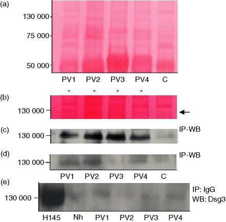
Pemphigus vulgaris immunoglobulin G can recognize a 130 000 MW antigen other than desmoglein 3 on peripheral blood mononuclear cell surface.
Cirillo N, Gombos F, Lanza A
Immunology 2007 Jul;121(3):377-82
Immunology 2007 Jul;121(3):377-82
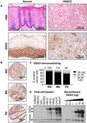
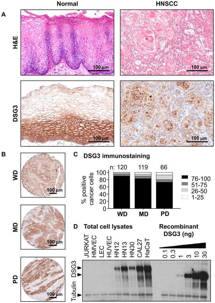
DSG3 is overexpressed in head neck cancer and is a potential molecular target for inhibition of oncogenesis.
Chen YJ, Chang JT, Lee L, Wang HM, Liao CT, Chiu CC, Chen PJ, Cheng AJ
Oncogene 2007 Jan 18;26(3):467-76
Oncogene 2007 Jan 18;26(3):467-76
Characterization of extracellular matrix components in the limbal epithelial stem cell compartment.
Schlötzer-Schrehardt U, Dietrich T, Saito K, Sorokin L, Sasaki T, Paulsson M, Kruse FE
Experimental eye research 2007 Dec;85(6):845-60
Experimental eye research 2007 Dec;85(6):845-60
New skin-equivalent model from de-epithelialized amnion membrane.
Yang L, Shirakata Y, Shudou M, Dai X, Tokumaru S, Hirakawa S, Sayama K, Hamuro J, Hashimoto K
Cell and tissue research 2006 Oct;326(1):69-77
Cell and tissue research 2006 Oct;326(1):69-77
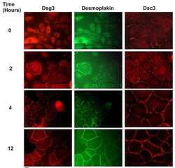
Desmoglein endocytosis and desmosome disassembly are coordinated responses to pemphigus autoantibodies.
Calkins CC, Setzer SV, Jennings JM, Summers S, Tsunoda K, Amagai M, Kowalczyk AP
The Journal of biological chemistry 2006 Mar 17;281(11):7623-34
The Journal of biological chemistry 2006 Mar 17;281(11):7623-34
Genetic and functional characterization of human pemphigus vulgaris monoclonal autoantibodies isolated by phage display.
Payne AS, Ishii K, Kacir S, Lin C, Li H, Hanakawa Y, Tsunoda K, Amagai M, Stanley JR, Siegel DL
The Journal of clinical investigation 2005 Apr;115(4):888-99
The Journal of clinical investigation 2005 Apr;115(4):888-99
Molecular classification of head and neck squamous cell carcinomas using patterns of gene expression.
Chung CH, Parker JS, Karaca G, Wu J, Funkhouser WK, Moore D, Butterfoss D, Xiang D, Zanation A, Yin X, Shockley WW, Weissler MC, Dressler LG, Shores CG, Yarbrough WG, Perou CM
Cancer cell 2004 May;5(5):489-500
Cancer cell 2004 May;5(5):489-500
Expression of desmosomal proteins in squamous cell carcinomas of the skin.
Kurzen H, Münzing I, Hartschuh W
Journal of cutaneous pathology 2003 Nov;30(10):621-30
Journal of cutaneous pathology 2003 Nov;30(10):621-30
Expression of desmosomal proteins in squamous cell carcinomas of the skin.
Kurzen H, Münzing I, Hartschuh W
Journal of cutaneous pathology 2003 Nov;30(10):621-30
Journal of cutaneous pathology 2003 Nov;30(10):621-30
No comments: Submit comment
Supportive validation
- Submitted by
- Invitrogen Antibodies (provider)
- Main image
- Experimental details
- NULL
- Submitted by
- Invitrogen Antibodies (provider)
- Main image

- Experimental details
- NULL

- Submitted by
- Invitrogen Antibodies (provider)
- Main image
- Experimental details
- NULL
- Submitted by
- Invitrogen Antibodies (provider)
- Main image
- Experimental details
- NULL
- Submitted by
- Invitrogen Antibodies (provider)
- Main image
- Experimental details
- NULL
- Submitted by
- Invitrogen Antibodies (provider)
- Main image
- Experimental details
- NULL
- Submitted by
- Invitrogen Antibodies (provider)
- Main image
- Experimental details
- NULL
- Submitted by
- Invitrogen Antibodies (provider)
- Main image
- Experimental details
- NULL
- Submitted by
- Invitrogen Antibodies (provider)
- Main image

- Experimental details
- NULL

- Submitted by
- Invitrogen Antibodies (provider)
- Main image
- Experimental details
- NULL

- Submitted by
- Invitrogen Antibodies (provider)
- Main image

- Experimental details
- NULL
- Submitted by
- Invitrogen Antibodies (provider)
- Main image
- Experimental details
- NULL
- Submitted by
- Invitrogen Antibodies (provider)
- Main image

- Experimental details
- NULL

- Submitted by
- Invitrogen Antibodies (provider)
- Main image
- Experimental details
- NULL
- Submitted by
- Invitrogen Antibodies (provider)
- Main image
- Experimental details
- NULL
- Submitted by
- Invitrogen Antibodies (provider)
- Main image
- Experimental details
- NULL
- Submitted by
- Invitrogen Antibodies (provider)
- Main image
- Experimental details
- NULL
- Submitted by
- Invitrogen Antibodies (provider)
- Main image
- Experimental details
- NULL
- Submitted by
- Invitrogen Antibodies (provider)
- Main image
- Experimental details
- NULL
- Submitted by
- Invitrogen Antibodies (provider)
- Main image

- Experimental details
- NULL
- Submitted by
- Invitrogen Antibodies (provider)
- Main image
- Experimental details
- NULL
- Submitted by
- Invitrogen Antibodies (provider)
- Main image
- Experimental details
- NULL

- Submitted by
- Invitrogen Antibodies (provider)
- Main image
- Experimental details
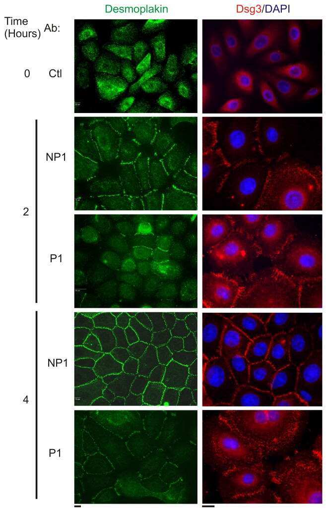
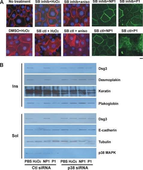
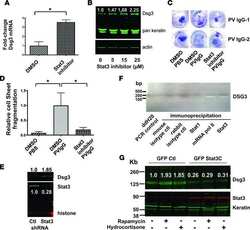
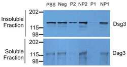
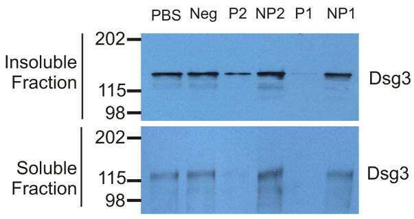
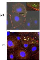
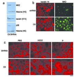
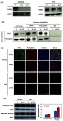
- Figure 4 Silencing of MK2 expression prevents PV mAb-induced loss of cell surface Dsg3 A) HaCat cells that stably express MK2 shRNA (shRNA) show markedly reduced MK2 and p38 protein levels compared to cells expressing control (Ctl) shRNA. Levels of histone H3 are shown as a loading control. B) Immunofluorescence staining of HaCat cells expressing MK2 or control (Ctl) shRNA confirms knockdown of MK2 expression 72 hours after transduction. C) shRNA silencing of MK2 expression decreases loss of cell surface Dsg3 (shown in red) 16 hours after treatment with pathogenic PV mAb (P) and oxidative stress (H 2 O 2 ). Scale bar=20mum.

- Submitted by
- Invitrogen Antibodies (provider)
- Main image
- Experimental details
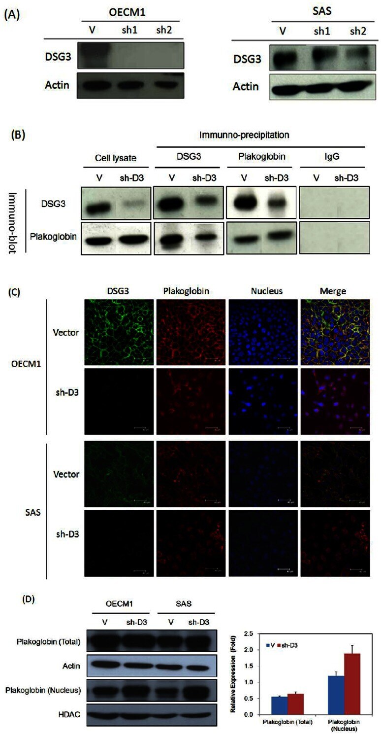
- Figure 1 DSG3 silencing disrupts the interaction of DSG3 with plakoglobin and induces plakoglobin nuclear translocation. ( A ) DSG3 expression was suppressed in shDSG3 cells using RNAi. OECM1 and SAS cells were transfected with the DSG3-specific shRNA expression plasmid or the empty vector plasmid, and clones were selected using G418. Four clones (OECM1-sh1 and OECM1-sh2 in OECM1 cells and SAS-sh1 and SAS-sh2 in SAS cells) of shDSG3 cells were chosen and analyzed using western blot assays to determine the DSG3 protein level. Two clones (OECM1-V in OECM1 cells and SAS-V in SAS cells) of the vector-transfected cells were also chosen as controls. The actin protein level was determined as an internal control for protein expression. ( B ) DSG3 silencing reduced the interaction of DSG3 with plakoglobin, as determined by immunoprecipitation (IP) and immunoblot (IB) analysis. Two sets of cells were examined, empty vector-transfected cells and shDSG3-transfected cells. In each sample, proteins were extracted and immunoprecipitated with anti-DSG3 (D3), anti-plakoglobin (Pg), mouse IgG (IgG) (as a negative control) as indicated above each lane. Each sample was subsequently immunoblotted with either a plakoglobin (Pg)- or DSG3 (D3)-specific antibody, as indicated on the left side of each set of samples. ( C ) DSG3 silencing induced plakoglobinn translocation from the cytoplasm to the nucleus. Immunofluorescence and confocal microscopy were used to examine the localization of DSG3 and pla
- Submitted by
- Invitrogen Antibodies (provider)
- Main image

- Experimental details
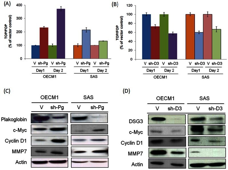
- Figure 3 DSG3 silencing suppresses the TCF/LEF transcriptional activity and the downstream target genes c-myc, cyclin D1, and MMP-7. ( A ) and ( B ) The effects of TCF/LEF transcriptional activity after Plakoglobin or DSG3 knockdown. The stable plakoglobin silencing (sh-Pg) or DSG3 silencing (sh-D3) cells were transfected with TOPflash or FOPflash luciferase reporter plasmid and the Renilla plasmid. After 24 hours, the luciferase activity was determined using the Steady-Glo Luciferase Reagent. The firefly luciferase activity was normalized against the Renilla luciferase activity and the fold increase of the TOPflash activity compared to the FOPflash activity was reported. ( C ) and ( D ) The expressions of the TCF/LEF downstream target genes c-myc and cyclin D1 was determined in the cells stably transfected with specific shRNAs target to plakoglobin (sh-Pg) or DSG3 (sh-D3) cells, compared to the vector transfected stably cells. In each sample, proteins were extracted and analyzed by western blot assays to determine the expression levels of c-myc, cyclin D1, and MMP-7.
- Submitted by
- Invitrogen Antibodies (provider)
- Main image

- Experimental details
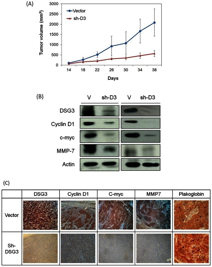
- Figure 5 DSG3 silencing suppressed the growth of xenografted tumors, which is associated with plakoglobin translocation and reduced expressions of TCF target genes. ( A ) Six of each group BALB/C mice were subcutaneously injected with 5x10 5 cells of the SAS cell line stably transfected with the vector (V) or the shDSG3 (sh2) plasmids. The tumor size was measured every 4 days starting two weeks after injection. Each dimension of the tumors were measured by calipers, and the tumor size was calculated as lengthxwidthxheight. (***: p
- Submitted by
- Invitrogen Antibodies (provider)
- Main image
- Experimental details
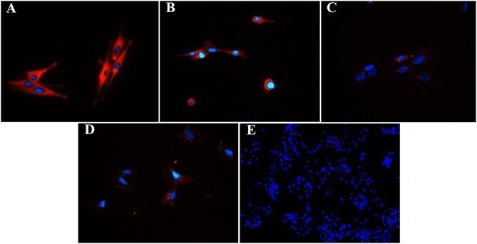
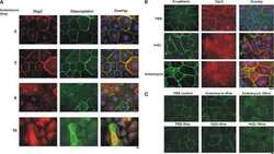
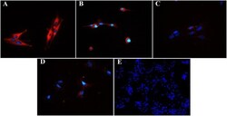
- Figure 8 Fluorescent photomicrographs of cultured progenitor cells (P3) from unaffected (A,B) and laminitic (C,D) hooves labeled (red) with anti- DSG1 (A,C) , DSG3 (B,D) antibodies or no antibodies (E) . DAPI nuclear stain (blue); Magnification = 40X (A-D) ; 20X (E) .