Explore
 Validate
 Learn
LearnPA5-96111
antibody from Invitrogen Antibodies
Targeting: H3-4
H3.4, H3/g, H3FT, H3t, HIST3H3
 Western blot
 ELISA
ELISA Immunocytochemistry Immunoprecipitation Immunohistochemistry Chromatin Immunoprecipitation
Antibody data
- Antibody Data
- Antigen structure
- References [0]
- Comments [0]
- Validations
- Immunocytochemistry [3]
- Immunoprecipitation [2]
- Immunohistochemistry [4]
- Chromatin Immunoprecipitation [2]
Submit
Validation data
Reference
Comment
Report error
- Product number
- PA5-96111 - Provider product page

- Provider
- Invitrogen Antibodies
- Product name
- Histone H3.1t Polyclonal Antibody
- Antibody type
- Polyclonal
- Antigen
- Synthetic peptide
- Description
- Positive Samples: Jurkat, A-431, Mouse liver, Mouse testis, Rat kidney; Cellular Location: Chromosome, Nucleus Immunogen sequence: VKKPHRYRPG TVALREIRRY QKSTELLIRK LPFQRLVREI AQDFKTDLRF QSSAVMALQE ACEAYLVGLF EDTNLCAIHA KRVTIMPKDI QLARRIRGER A
- Reactivity
- Human, Mouse, Rat
- Host
- Rabbit
- Isotype
- IgG
- Vial size
- 100 μL
- Concentration
- 2.17 mg/mL
- Storage
- -20°C, Avoid Freeze/Thaw Cycles
No comments: Submit comment
Supportive validation
- Submitted by
- Invitrogen Antibodies (provider)
- Main image
- Experimental details
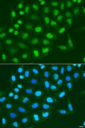
- Immunocytochemistry-Immunofluorescence analysis of Histone H3.1t was performed in U2OS cells using Histone H3.1t Polyclonal Antibody (Product # PA5-96111).
- Submitted by
- Invitrogen Antibodies (provider)
- Main image
- Experimental details
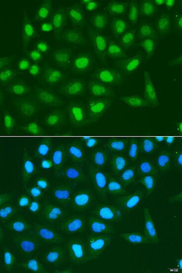
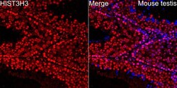
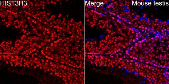
- Immunofluorescence analysis of Histone H3.1t in mouse testis cells. Samples were incubated with Histone H3.1t Polyclonal antibody (Product # PA5-96111) using a dilution of 1:200 (40x lens). Blue: DAPI for nuclear staining.
- Submitted by
- Invitrogen Antibodies (provider)
- Main image

- Experimental details
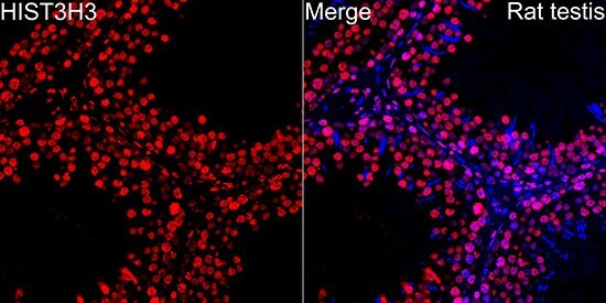
- Immunofluorescence analysis of Histone H3.1t in rat testis cells. Samples were incubated with Histone H3.1t Polyclonal antibody (Product # PA5-96111) using a dilution of 1:200 (40x lens). Blue: DAPI for nuclear staining.
Supportive validation
- Submitted by
- Invitrogen Antibodies (provider)
- Main image
- Experimental details
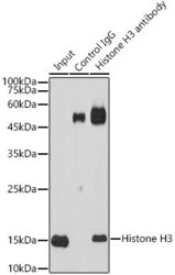
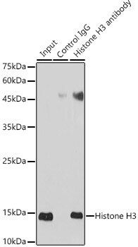
- Immunoprecipitation of Histone H3.1t in 600 μg extracts of HeLa cells. Samples were precipitated with 10 μg Histone H3.1t Polyclonal antibody (Product # PA5-96111). Western blot was performed from the immunoprecipitate using Histone H3.1t Polyclonal antibody (Product # PA5-96111) at a dilution of 1:30,000.

- Submitted by
- Invitrogen Antibodies (provider)
- Main image
- Experimental details
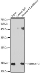
- Immunoprecipitation of Histone H3.1t in 600 μg extracts of HeLa cells. Samples were precipitated with 10 μg Histone H3.1t Polyclonal antibody (Product # PA5-96111). Western blot was performed from the immunoprecipitate using Histone H3.1t Polyclonal antibody (Product # PA5-96111) at a dilution of 1:30,000.
Supportive validation
- Submitted by
- Invitrogen Antibodies (provider)
- Main image
- Experimental details
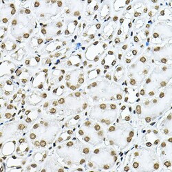
- Immunohistochemistry analysis of Histone H3.1t in paraffin-embedded mouse kidney. Samples were incubated with Histone H3.1t Polyclonal antibody (Product # PA5-96111) using a dilution of 1:100 (40x lens). Perform high pressure antigen retrieval with 10 mM citrate buffer pH 6.0 before commencing with IHC staining protocol.
- Submitted by
- Invitrogen Antibodies (provider)
- Main image
- Experimental details
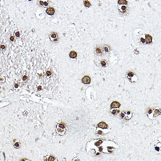
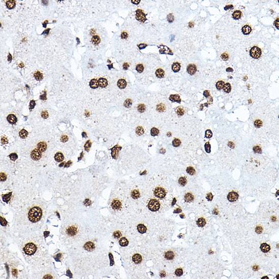
- Immunohistochemistry analysis of Histone H3.1t in paraffin-embedded human brain. Samples were incubated with Histone H3.1t Polyclonal antibody (Product # PA5-96111) using a dilution of 1:100 (40x lens). Perform high pressure antigen retrieval with 10 mM citrate buffer pH 6.0 before commencing with IHC staining protocol.
- Submitted by
- Invitrogen Antibodies (provider)
- Main image
- Experimental details
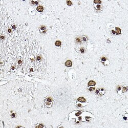
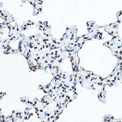
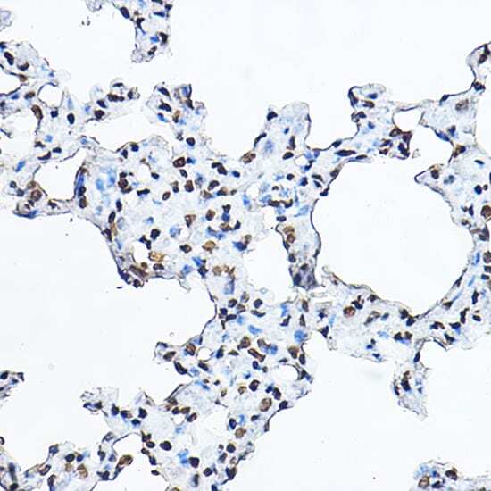
- Immunohistochemistry analysis of Histone H3.1t in paraffin-embedded rat lung. Samples were incubated with Histone H3.1t Polyclonal antibody (Product # PA5-96111) using a dilution of 1:100 (40x lens). Perform high pressure antigen retrieval with 10 mM citrate buffer pH 6.0 before commencing with IHC staining protocol.
- Submitted by
- Invitrogen Antibodies (provider)
- Main image
- Experimental details
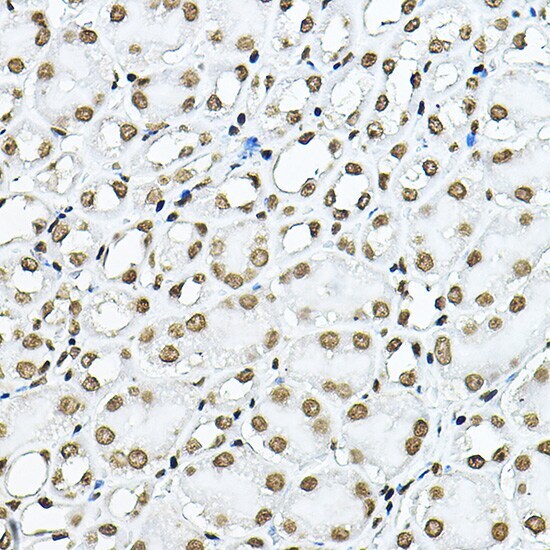
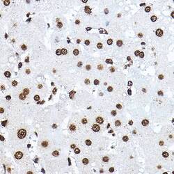
- Immunohistochemistry analysis of Histone H3.1t in paraffin-embedded human liver. Samples were incubated with Histone H3.1t Polyclonal antibody (Product # PA5-96111) using a dilution of 1:100 (40x lens). Perform high pressure antigen retrieval with 10 mM citrate buffer pH 6.0 before commencing with IHC staining protocol.
Supportive validation
- Submitted by
- Invitrogen Antibodies (provider)
- Main image

- Experimental details
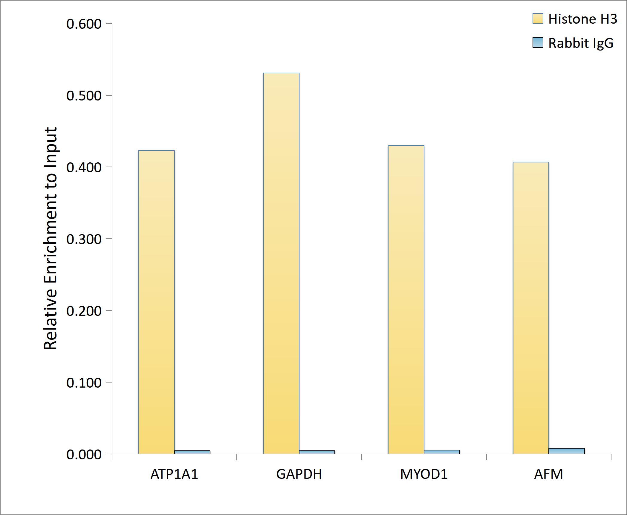
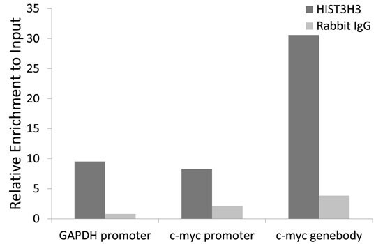
- Chromatin immunoprecipitation analysis of Histone H3.1t in HeLa cell line using Histone H3.1t Polyclonal Antibody (Product # PA5-96111) and rabbit IgG. The amount of immunoprecipitated DNA was checked by quantitative PCR. Histogram was constructed by the ratios of the immunoprecipitated DNA to the input.
- Submitted by
- Invitrogen Antibodies (provider)
- Main image

- Experimental details
- ChIP analysis of Histone H3.1t in extracts of 293T cells. Samples were incubated with Histone H3.1t Polyclonal antibody (Product # PA5-96111) and rabbit IgG. The amount of immunoprecipitated DNA was checked by quantitative PCR. Histogram was constructed by the ratios of the immunoprecipitated DNA to the input.