Explore
 Validate
 Learn
Learn Western blot
 Immunohistochemistry
Antibody data
- Antibody Data
- Antigen structure
- References [1]
- Comments [0]
- Validations
- Western blot [1]
Submit
Validation data
Reference
Comment
Report error
- Product number
- PA2023-1 - Provider product page

- Provider
- Boster Biological Technology
- Product name
- Anti-DISC1 Antibody
- Antibody type
- Polyclonal
- Description
- Polyclonal antibody for DISC1 detection. Host: Rabbit.Size: 100μg/vial. Tested applications: IHC-P. Reactive species: Human. DISC1 information: Molecular Weight: 93611 MW; Subcellular Localization: Cytoplasm. Cytoplasm, cytoskeleton. Cytoplasm, cytoskeleton, microtubule organizing center, centrosome. Cell junction, synapse, postsynaptic cell membrane, postsynaptic density . Colocalizes with NDEL1 in the perinuclear region and the centrosome (By similarity). Localizes to punctate cytoplasmic foci which overlap in part with mitochondria. Colocalizes with PCNT at the centrosome; Tissue Specificity: Ubiquitous. Highly expressed in the dentate gyrus of the hippocampus. Also expressed in the temporal and parahippocampal cortices and cells of the white matter.
- Reactivity
- Human, Mouse, Rat
- Host
- Rabbit
- Vial size
- 100μg/vial
- Concentration
- Add 0.2ml of distilled water will yield a concentration of 500ug/ml.
- Storage
- At -20°C for one year. After reconstitution, at 4°C for one month. It can also be aliquoted and stored frozen at -20°C for a longer time. Avoid repeated freezing and thawing.
- Handling
- Add 0.2ml of distilled water will yield a concentration of 500ug/ml.
Submitted references Association of Mental Health-Related Proteins DAXX, DRD3, and DISC1 With the Progression and Prognosis of Chondrosarcoma.
He L, Shi X, Chen R, Wu Z, Yang Z, Li Z
Frontiers in molecular biosciences 2019;6:134
Frontiers in molecular biosciences 2019;6:134
No comments: Submit comment
Supportive validation
- Submitted by
- Boster Biological Technology (provider)
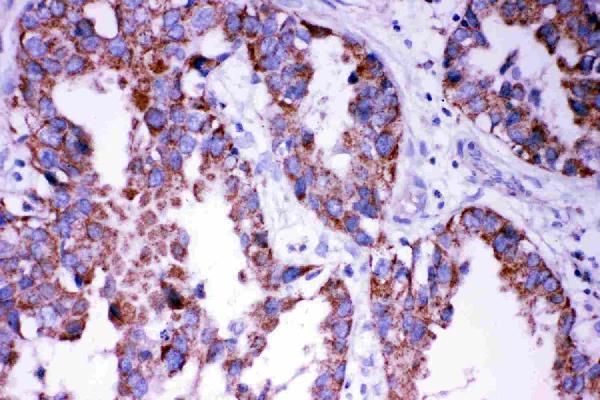
- Main image
- Experimental details
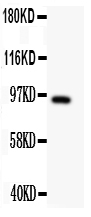
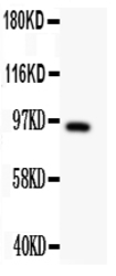
- Western blot analysis of DISC1 using anti- DISC1 antibody (PA2023-1). Electrophoresis was performed on a 5-20% SDS-PAGE gel at 70V (Stacking gel) / 90V (Resolving gel) for 2-3 hours. The sample well of each lane was loaded with 50ug of sample under reducing conditions. Lane 1: U87 Whole Cell Lysate. After Electrophoresis, proteins were transferred to a Nitrocellulose membrane at 150mA for 50-90 minutes. Blocked the membrane with 5% Non-fat Milk/ TBS for 1.5 hour at RT. The membrane was incubated with rabbit anti- DISC1antigen affinity purified polyclonal antibody (Catalog # PA2023-1) at 0.5 μg/mL overnight at 4°C, then washed with TBS-0.1%Tween 3 times with 5 minutes each and probed with a goat anti-rabbit IgG-HRP secondary antibody at a dilution of 1:10000 for 1.5 hour at RT. The signal is developed using an Enhanced Chemiluminescent detection (ECL) kit (Catalog # EK1002) with Tanon 5200 system. A specific band was detected for DISC1 at approximately 94KD. The expected band size for DISC1 is at 94KD.
- Additional image