Explore
 Validate
 Learn
Learn Western blot
 Immunocytochemistry
 Immunohistochemistry
Antibody data
- Antibody Data
- Antigen structure
- References [3]
- Comments [0]
- Validations
- Immunocytochemistry [2]
- Flow cytometry [1]
- Other assay [1]
Submit
Validation data
Reference
Comment
Report error
- Product number
- 40-6900 - Provider product page

- Provider
- Invitrogen Antibodies
- Product name
- DISC1 Polyclonal Antibody
- Antibody type
- Polyclonal
- Antigen
- Synthetic peptide
- Reactivity
- Human, Mouse, Rat
- Host
- Rabbit
- Isotype
- IgG
- Vial size
- 100 μg
- Concentration
- 0.25 mg/mL
- Storage
- -20°C
Submitted references An inherited duplication at the gene p21 Protein-Activated Kinase 7 (PAK7) is a risk factor for psychosis.
DISC1 regulates primary cilia that display specific dopamine receptors.
Disc1 regulates granule cell migration in the developing hippocampus.
Morris DW, Pearson RD, Cormican P, Kenny EM, O'Dushlaine CT, Perreault LP, Giannoulatou E, Tropea D, Maher BS, Wormley B, Kelleher E, Fahey C, Molinos I, Bellini S, Pirinen M, Strange A, Freeman C, Thiselton DL, Elves RL, Regan R, Ennis S, Dinan TG, McDonald C, Murphy KC, O'Callaghan E, Waddington JL, Walsh D, O'Donovan M, Grozeva D, Craddock N, Stone J, Scolnick E, Purcell S, Sklar P, Coe B, Eichler EE, Ophoff R, Buizer J, Szatkiewicz J, Hultman C, Sullivan P, Gurling H, Mcquillin A, St Clair D, Rees E, Kirov G, Walters J, Blackwood D, Johnstone M, Donohoe G, International Schizophrenia Consortium, SGENE+ Consortium, O'Neill FA, Wellcome Trust Case Control Consortium 2, Kendler KS, Gill M, Riley BP, Spencer CC, Corvin A
Human molecular genetics 2014 Jun 15;23(12):3316-26
Human molecular genetics 2014 Jun 15;23(12):3316-26
DISC1 regulates primary cilia that display specific dopamine receptors.
Marley A, von Zastrow M
PloS one 2010 May 28;5(5):e10902
PloS one 2010 May 28;5(5):e10902
Disc1 regulates granule cell migration in the developing hippocampus.
Meyer KD, Morris JA
Human molecular genetics 2009 Sep 1;18(17):3286-97
Human molecular genetics 2009 Sep 1;18(17):3286-97
No comments: Submit comment
Supportive validation
- Submitted by
- Invitrogen Antibodies (provider)
- Main image
- Experimental details
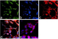
- Immunofluorescence analysis of DISC1 was done on 70% confluent log phase SH-SY5Y cells. The cells were fixed with 4% paraformaldehyde for 15 minutes, permeabilized with 0.25% Triton™ X-100 for 10 minutes, and blocked with 5% BSA for 1 hour at room temperature. The cells were labeled with DISC1 Rabbit Polyclonal Antibody (Product # 40-6900) at 1 µg/mL in 1% BSA and incubated for 3 hours at room temperature and then labeled with Goat anti-Rabbit IgG (H+L) Superclonal™ Secondary Antibody, Alexa Fluor® 488 conjugate (Product # A27034) at a dilution of 1:2000 for 45 minutes at room temperature (Panel a: green). Nuclei (Panel b: blue) were stained with SlowFade® Gold Antifade Mountant with DAPI (Product # S36938). F-actin (Panel c: red) was stained with Alexa Fluor® 555 Rhodamine Phalloidin (Product # R415, 1:300). Panel d is a merged image showing Nuclear localization. Panel e is a no primary antibody control. The images were captured at 60X magnification.

- Submitted by
- Invitrogen Antibodies (provider)
- Main image

- Experimental details
- Immunofluorescence analysis of DISC1 was done on 70% confluent log phase SH-SY5Y cells. The cells were fixed with 4% paraformaldehyde for 15 minutes, permeabilized with 0.25% Triton™ X-100 for 10 minutes, and blocked with 5% BSA for 1 hour at room temperature. The cells were labeled with DISC1 Rabbit Polyclonal Antibody (Product # 40-6900) at 1 µg/mL in 1% BSA and incubated for 3 hours at room temperature and then labeled with Goat anti-Rabbit IgG (Heavy Chain) Superclonal™ Secondary Antibody, Alexa Fluor® 488 conjugate (Product # A27034) at a dilution of 1:2000 for 45 minutes at room temperature (Panel a: green). Nuclei (Panel b: blue) were stained with SlowFade® Gold Antifade Mountant with DAPI (Product # S36938). F-actin (Panel c: red) was stained with Alexa Fluor® 555 Rhodamine Phalloidin (Product # R415, 1:300). Panel d is a merged image showing Nuclear localization. Panel e is a no primary antibody control. The images were captured at 60X magnification.

Supportive validation
- Submitted by
- Invitrogen Antibodies (provider)
- Main image

- Experimental details
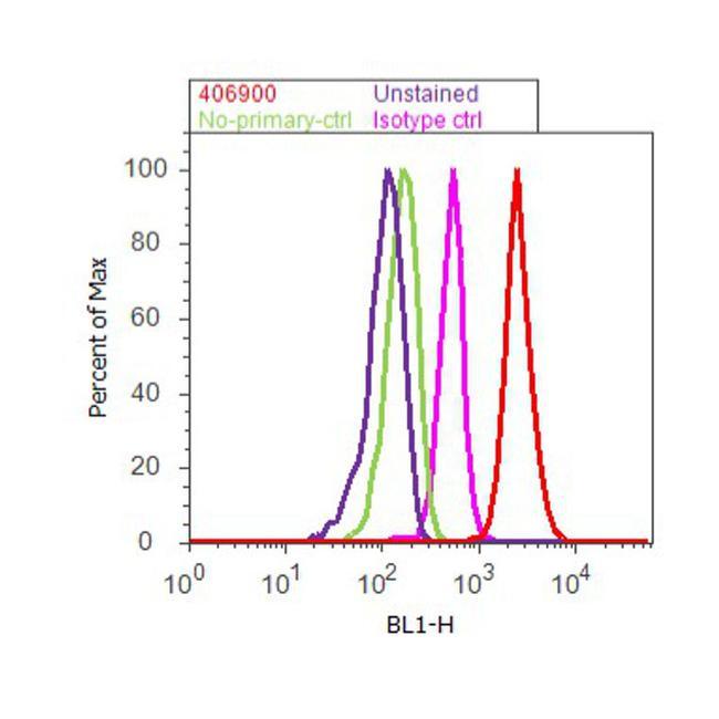
- Flow cytometry analysis of DISC1 was done on SH-SY5Y cells. Cells were fixed with 70% ethanol for 10 minutes, permeabilized with 0.25% Triton™ X-100 for 20 minutes, and blocked with 5% BSA for 30 minutes at room temperature. Cells were labeled with DISC1 Rabbit Polyclonal Antibody (406900, red histogram) or with rabbit isotype control (pink histogram) at 3-5 ug/million cells in 2.5% BSA. After incubation at room temperature for 2 hours, the cells were labeled with Alexa Fluor® 488 Goat Anti-Rabbit Secondary Antibody (A11008) at a dilution of 1:400 for 30 minutes at room temperature. The representative 10,000 cells were acquired and analyzed for each sample using an Attune® Acoustic Focusing Cytometer. The purple histogram represents unstained control cells and the green histogram represents no-primary-antibody control.
Supportive validation
- Submitted by
- Invitrogen Antibodies (provider)
- Main image
- Experimental details
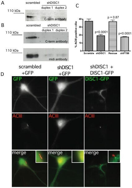
- Figure 7 DISC1 knockdown and rescue in striatal neurons. ( A ) Example immunoblot showing depletion of endogenous DISC1 detected in PC12 cells using the C-terminal antibody (see Materials and Methods ). The immunoreactive band corresponding to the major full length DISC1 species is shown. ( B ) Example of DISC1 depletion in striatal cultures, detected using the C-terminal (C-term) antibody and verified using the midi antibody, as indicated. ( C ) Quantification of knockdown and rescue phenotypes, based on count of cilia number compiled from 8 independent experiments. ( D ) Examples of the phenotypes observed in the indicated conditions (indicated above each column). The rescue condition (right column of images) shows the localization of DISC1-GFP rather than GFP. Insets indicate relevant regions of the cell body at higher magnification.