Explore
 Validate
 Learn
Learn Western blot
 ELISA
ELISAAntibody data
- Antibody Data
- Antigen structure
- References [12]
- Comments [0]
- Validations
- Western blot [2]
- Immunocytochemistry [1]
Submit
Validation data
Reference
Comment
Report error
- Product number
- 13471-1-AP - Provider product page

- Provider
- Proteintech Group
- Proper citation
- Proteintech Cat#13471-1-AP, RRID:AB_2183417
- Product name
- SDCCAG8 antibody
- Antibody type
- Polyclonal
- Description
- KD/KO validated SDCCAG8 antibody (Cat. #13471-1-AP) is a rabbit polyclonal antibody that shows reactivity with human, mouse, rat, zebrafish, Canine and has been validated for the following applications: IF, IHC, WB,ELISA.
- Reactivity
- Human, Mouse, Rat, Canine, Zebrafish
- Host
- Rabbit
- Conjugate
- Unconjugated
- Isotype
- IgG
- Vial size
- 20ul, 150ul
Submitted references Loss of C-Terminal Coiled-Coil Domains in SDCCAG8 Impairs Centriolar Satellites and Causes Defective Sperm Flagellum Biogenesis and Male Fertility.
Characterization of two novel knock-in mouse models of syndromic retinal ciliopathy carrying hypomorphic Sdccag8 mutations.
The carboxyl-terminal region of SDCCAG8 comprises a functional module essential for cilia formation as well as organ development and homeostasis.
Altered gene regulation as a candidate mechanism by which ciliopathy gene SDCCAG8 contributes to schizophrenia and cognitive function.
The Role of the Microglial Cx3cr1 Pathway in the Postnatal Maturation of Retinal Photoreceptors.
Centrobin controls primary ciliogenesis in vertebrates.
Cby1 promotes Ahi1 recruitment to a ring-shaped domain at the centriole-cilium interface and facilitates proper cilium formation and function.
Renal-retinal ciliopathy gene Sdccag8 regulates DNA damage response signaling.
SDCCAG8 regulates pericentriolar material recruitment and neuronal migration in the developing cortex.
Selective loss of RPGRIP1-dependent ciliary targeting of NPHP4, RPGR and SDCCAG8 underlies the degeneration of photoreceptor neurons.
Global landscape of HIV-human protein complexes.
Candidate exome capture identifies mutation of SDCCAG8 as the cause of a retinal-renal ciliopathy.
Li K, Zhou X, Liu W, Wang Y, Zhang Z, Zhang H, Jiang L
Cells 2025 Jul 23;14(15)
Cells 2025 Jul 23;14(15)
Characterization of two novel knock-in mouse models of syndromic retinal ciliopathy carrying hypomorphic Sdccag8 mutations.
Ren ZL, Zhang HB, Li L, Yang ZL, Jiang L
Zoological research 2022 May 18;43(3):442-456
Zoological research 2022 May 18;43(3):442-456
The carboxyl-terminal region of SDCCAG8 comprises a functional module essential for cilia formation as well as organ development and homeostasis.
Tsutsumi R, Chaya T, Tsujii T, Furukawa T
The Journal of biological chemistry 2022 Mar;298(3):101686
The Journal of biological chemistry 2022 Mar;298(3):101686
Altered gene regulation as a candidate mechanism by which ciliopathy gene SDCCAG8 contributes to schizophrenia and cognitive function.
Flynn M, Whitton L, Donohoe G, Morrison CG, Morris DW
Human molecular genetics 2020 Feb 1;29(3):407-417
Human molecular genetics 2020 Feb 1;29(3):407-417
The Role of the Microglial Cx3cr1 Pathway in the Postnatal Maturation of Retinal Photoreceptors.
Jobling AI, Waugh M, Vessey KA, Phipps JA, Trogrlic L, Greferath U, Mills SA, Tan ZL, Ward MM, Fletcher EL
The Journal of neuroscience : the official journal of the Society for Neuroscience 2018 May 16;38(20):4708-4723
The Journal of neuroscience : the official journal of the Society for Neuroscience 2018 May 16;38(20):4708-4723
Centrobin controls primary ciliogenesis in vertebrates.
Ogungbenro YA, Tena TC, Gaboriau D, Lalor P, Dockery P, Philipp M, Morrison CG
The Journal of cell biology 2018 Apr 2;217(4):1205-1215
The Journal of cell biology 2018 Apr 2;217(4):1205-1215
Cby1 promotes Ahi1 recruitment to a ring-shaped domain at the centriole-cilium interface and facilitates proper cilium formation and function.
Lee YL, Santé J, Comerci CJ, Cyge B, Menezes LF, Li FQ, Germino GG, Moerner WE, Takemaru K, Stearns T
Molecular biology of the cell 2014 Oct 1;25(19):2919-33
Molecular biology of the cell 2014 Oct 1;25(19):2919-33
Renal-retinal ciliopathy gene Sdccag8 regulates DNA damage response signaling.
Airik R, Slaats GG, Guo Z, Weiss AC, Khan N, Ghosh A, Hurd TW, Bekker-Jensen S, Schrøder JM, Elledge SJ, Andersen JS, Kispert A, Castelli M, Boletta A, Giles RH, Hildebrandt F
Journal of the American Society of Nephrology : JASN 2014 Nov;25(11):2573-83
Journal of the American Society of Nephrology : JASN 2014 Nov;25(11):2573-83
SDCCAG8 regulates pericentriolar material recruitment and neuronal migration in the developing cortex.
Insolera R, Shao W, Airik R, Hildebrandt F, Shi SH
Neuron 2014 Aug 20;83(4):805-22
Neuron 2014 Aug 20;83(4):805-22
Selective loss of RPGRIP1-dependent ciliary targeting of NPHP4, RPGR and SDCCAG8 underlies the degeneration of photoreceptor neurons.
Patil H, Tserentsoodol N, Saha A, Hao Y, Webb M, Ferreira PA
Cell death & disease 2012 Jul 19;3(7):e355
Cell death & disease 2012 Jul 19;3(7):e355
Global landscape of HIV-human protein complexes.
Jäger S, Cimermancic P, Gulbahce N, Johnson JR, McGovern KE, Clarke SC, Shales M, Mercenne G, Pache L, Li K, Hernandez H, Jang GM, Roth SL, Akiva E, Marlett J, Stephens M, D'Orso I, Fernandes J, Fahey M, Mahon C, O'Donoghue AJ, Todorovic A, Morris JH, Maltby DA, Alber T, Cagney G, Bushman FD, Young JA, Chanda SK, Sundquist WI, Kortemme T, Hernandez RD, Craik CS, Burlingame A, Sali A, Frankel AD, Krogan NJ
Nature 2011 Dec 21;481(7381):365-70
Nature 2011 Dec 21;481(7381):365-70
Candidate exome capture identifies mutation of SDCCAG8 as the cause of a retinal-renal ciliopathy.
Otto EA, Hurd TW, Airik R, Chaki M, Zhou W, Stoetzel C, Patil SB, Levy S, Ghosh AK, Murga-Zamalloa CA, van Reeuwijk J, Letteboer SJ, Sang L, Giles RH, Liu Q, Coene KL, Estrada-Cuzcano A, Collin RW, McLaughlin HM, Held S, Kasanuki JM, Ramaswami G, Conte J, Lopez I, Washburn J, Macdonald J, Hu J, Yamashita Y, Maher ER, Guay-Woodford LM, Neumann HP, Obermüller N, Koenekoop RK, Bergmann C, Bei X, Lewis RA, Katsanis N, Lopes V, Williams DS, Lyons RH, Dang CV, Brito DA, Dias MB, Zhang X, Cavalcoli JD, Nürnberg G, Nürnberg P, Pierce EA, Jackson PK, Antignac C, Saunier S, Roepman R, Dollfus H, Khanna H, Hildebrandt F
Nature genetics 2010 Oct;42(10):840-50
Nature genetics 2010 Oct;42(10):840-50
No comments: Submit comment
Supportive validation
- Submitted by
- Proteintech Group (provider)
- Main image
- Experimental details
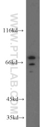
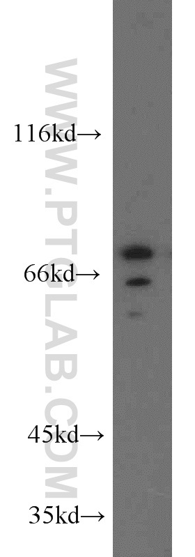
- PC-3 cells were subjected to SDS PAGE followed by western blot with 13471-1-AP(SDCCAG8 antibody) at dilution of 1:500
- Sample type
- cell line
- Submitted by
- Proteintech Group (provider)
- Main image
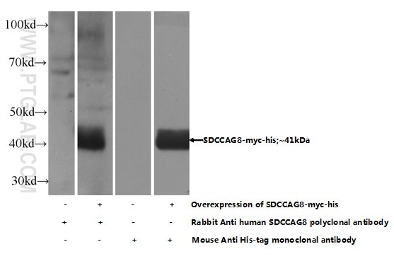
- Experimental details
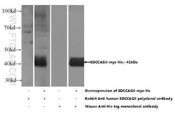
- Transfected HEK-293 cells were subjected to SDS PAGE followed by western blot with 13471-1-AP( SDCCAG8 Antibody) at dilution of 1:500
- Sample type
- cell line
Supportive validation
- Submitted by
- Proteintech Group (provider)
- Main image
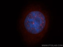
- Experimental details
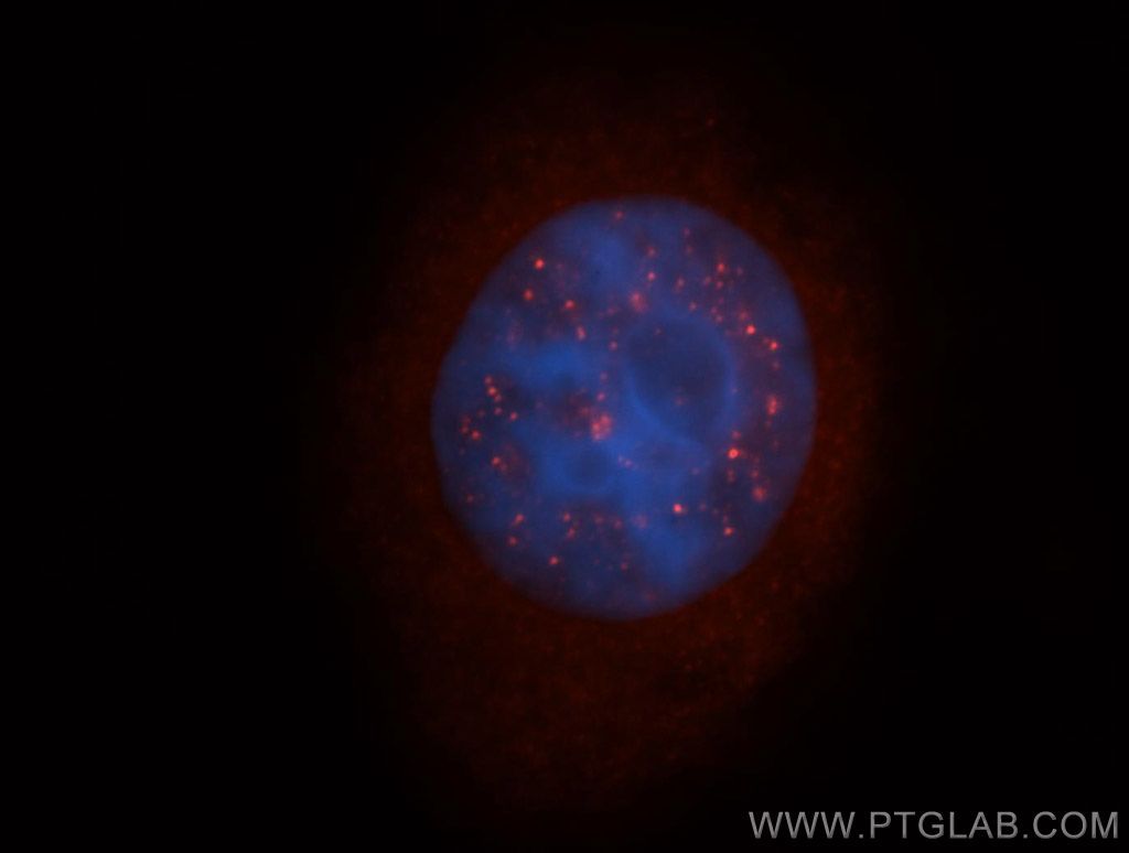
- Immunofluorescent analysis of Hela cells, using SDCCAG8 antibody 13471-1-AP at 1:50 dilution and Rhodamine-labeled goat anti-rabbit IgG (red). Blue pseudocolor = DAPI (fluorescent DNA dye).
- Sample type
- cell line