Explore
 Validate
 Learn
Learn Western blot
 Immunocytochemistry
Antibody data
- Antibody Data
- Antigen structure
- References [1]
- Comments [0]
- Validations
- Immunocytochemistry [1]
- Immunohistochemistry [1]
Submit
Validation data
Reference
Comment
Report error
- Product number
- GTX15776 - Provider product page

- Provider
- GeneTex
- Proper citation
- GeneTex Cat#GTX15776, RRID:AB_2687516
- Product name
- Neurofibromin antibody [McNFn27b]
- Antibody type
- Monoclonal
- Reactivity
- Human, Mouse, Rat
- Host
- Mouse
Submitted references Tumor Evolution of Glioma-Intrinsic Gene Expression Subtypes Associates with Immunological Changes in the Microenvironment.
Wang Q, Hu B, Hu X, Kim H, Squatrito M, Scarpace L, deCarvalho AC, Lyu S, Li P, Li Y, Barthel F, Cho HJ, Lin YH, Satani N, Martinez-Ledesma E, Zheng S, Chang E, Sauvé CG, Olar A, Lan ZD, Finocchiaro G, Phillips JJ, Berger MS, Gabrusiewicz KR, Wang G, Eskilsson E, Hu J, Mikkelsen T, DePinho RA, Muller F, Heimberger AB, Sulman EP, Nam DH, Verhaak RGW
Cancer cell 2017 Jul 10;32(1):42-56.e6
Cancer cell 2017 Jul 10;32(1):42-56.e6
No comments: Submit comment
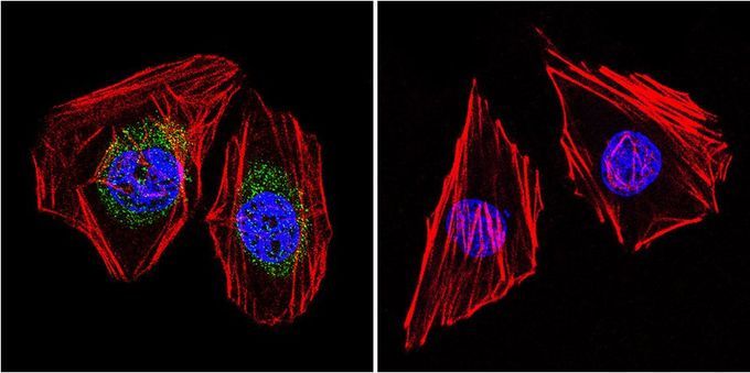
Supportive validation
- Submitted by
- GeneTex (provider)
- Main image
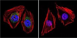
- Experimental details
- Immunofluorescent analysis of Neurofibromin in HeLa cells. Cells were grown on slides and fixed with formaldehyde prior to staining. Cells were probed without (control) or with Neurofibromin antibody [McNFn27b] at a dilution of 1:20 overnight at 4¢XC, washed with PBS and incubated with a proper secondary antibody. Neurofibromin staining (green), F-Actin staining with Phalloidin (red) and nuclei with DAPI (blue) is shown. Images were taken at 60X magnification.
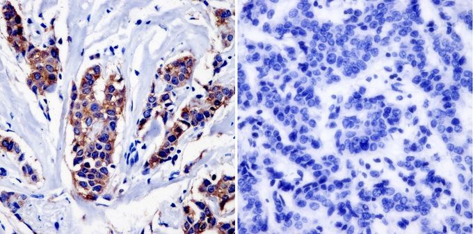
Supportive validation
- Submitted by
- GeneTex (provider)
- Main image
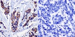
- Experimental details
- Immunohistochemistry was performed on cancer biopsies of deparaffinized human breast carcinoma tissue. To expose target proteins, heat induced antigen retrieval was performed using 10mM sodium citrate (pH6.0) buffer, microwaved for 8-15 minutes. Following antigen retrieval tissues were blocked in 3% BSA-PBS for 30 minutes at room temperature and probed at a dilution of 1:20 with or without Neurofibromin antibody [McNFn27b] overnight at 4¢XC in a humidified chamber. Tissues were washed with PBST and endogenous peroxidase activity was quenched with a peroxidase suppressor. Detection was performed using a biotin-conjugated secondary antibody and SA-HRP, followed by colorimetric detection using DAB. Tissues were counterstained with hematoxylin and prepped for mounting.