Explore
 Validate
 Learn
Learn Western blot
 ELISA
ELISAAntibody data
- Antibody Data
- Antigen structure
- References [1]
- Comments [0]
- Validations
- Western blot [1]
- Immunohistochemistry [1]
Submit
Validation data
Reference
Comment
Report error
- Product number
- NB300-153 - Provider product page

- Provider
- Novus Biologicals
- Proper citation
- Novus Cat#NB300-153, RRID:AB_10003032
- Product name
- Mouse Monoclonal Neurofibromin 1 Antibody
- Antibody type
- Monoclonal
- Description
- Protein A or G purified. This is specific for full-length or truncated forms of neurofibromin.
- Reactivity
- Human, Mouse, Rat, Guinea Pig
- Host
- Mouse
- Isotype
- IgG
- Vial size
- 0.2 ml
- Concentration
- 1 mg/ml
- Storage
- Store at 4C short term. Aliquot and store at -20C long term. Avoid freeze-thaw cycles.
Submitted references S100B and neurofibromin immunostaining and X-inactivation patterns of laser-microdissected cells indicate a multicellular origin of some NF1-associated neurofibromas.
Tucker T, Riccardi VM, Brown C, Fee J, Sutcliffe M, Vielkind J, Wechsler J, Wolkenstein P, Friedman JM
Journal of neuroscience research 2011 Sep;89(9):1451-60
Journal of neuroscience research 2011 Sep;89(9):1451-60
No comments: Submit comment
Supportive validation
- Submitted by
- Novus Biologicals (provider)
- Main image
- Experimental details
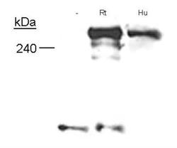
- Western Blot: Neurofibromin 1 Antibody (McNFn27a) [NB300-153] - Specificity of Neurofibromin, using NB 300-153, in Schwann cells from embryonic knockout mice, adult human nerves and neonatal rat nerves.

Supportive validation
- Submitted by
- Novus Biologicals (provider)
- Main image
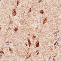
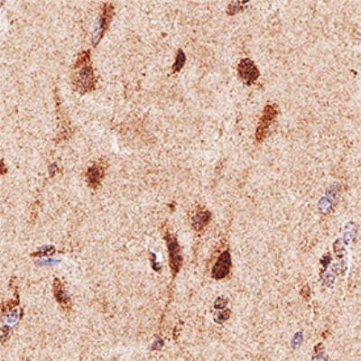
- Experimental details
- Immunohistochemistry-Paraffin: Neurofibromin 1 Antibody (McNFn27a) [NB300-153] - IHC analysis of formalin fixed paraffin-embedded (FFPE) human brain using Neurofibromin 1 antibody at 1:50 on a Bond Rx autostainer (Leica Biosystems). The assay involved 20 minutes of heat induced antigen retrieval (HIER) using 10mM sodium citrate buffer (pH 6.0) and endogenous peroxidase quenching with peroxide block. The sections were incubated with primary antibody for 30 minutes and Bond Polymer Refine Detection (Leica Biosystems) with DAB was used for signal development followed by counterstaining with hematoxylin. Whole slide scanning and capturing of representative images was performed using Aperio AT2 (Leica Biosystems). Nuclear staining of Neurofibromin 1was observed. Staining was performed by Histowiz.